Our Building Blocks
Atoms
Atoms are the smallest building blocks of all matter. Not just in biology, in everything. Iron, oxygen, carbon, copper, these are all atoms. The word atom comes from the Greek word atomos, which means “undivided” or “uncut.”
Every atom has three parts:
- Protons: positively charged particles in the centre (nucleus)
- Neutrons: neutral particles also in the centre
- Electrons: negatively charged particles that orbit around the outside
Electrons want to exist in pairs. Each orbital holds exactly 2 electrons. When an atom or molecule has an unpaired electron, it becomes chemically unstable and will aggressively try to steal an electron from the nearest molecule to re-stabilise itself. A molecule in this unstable state is called a free radical.
Molecules
A molecule is two or more atoms held together by chemical bonds. Water is a molecule (two hydrogen atoms bonded to one oxygen). So is glucose, cholesterol, oestrogen, dopamine, and every protein in your body.
Some molecules are small enough to pass through cell membranes freely (oxygen, carbon dioxide, water, fat-soluble hormones like testosterone). Others are too large or too charged and need transporter proteins to get them across (glucose, amino acids, water-soluble hormones like insulin). This is why steroid hormones act inside the cell on DNA while peptide hormones act on receptors on the outside of the cell.
DNA
DNA is a very long molecule made of a sequence of smaller molecules called nucleotides. Each nucleotide contains a sugar, a phosphate group, and one of four chemical bases: adenine (A), thymine (T), cytosine (C), and guanine (G). The sequence of these four bases is the genetic code. Your DNA is the instruction manual for making you, divided into segments called chromosomes and shorter individual units called genes. The sum of all your genes is the genome.
What genes specifically do is provide instructions for building proteins. Most of the functional machinery in your body is made of protein, including enzymes (which speed up chemical reactions), hormones (which carry chemical messages), antibodies (which fight infection), and structural materials (like collagen). Your genome contains roughly 20,000-25,000 protein-coding genes.
Gene expression
Every single cell in your body contains a complete copy of all 20,000+ genes. A muscle cell has the same DNA as a liver cell, a brain cell, or a skin cell. What makes them different isn't the genes they have, it's which genes they actively read.
- A muscle cell reads the genes for contractile proteins like actin and myosin
- A liver cell reads the genes for SHBG, albumin, and clotting factors
- A skin fibroblast reads the genes for collagen and elastin
- A pancreatic beta cell reads the gene for insulin
The same complete instruction manual sits in every cell, but each cell type only opens the chapters relevant to its job. This is called gene expression, which genes are switched on (expressed) and which are switched off (silenced) in a given cell at a given moment.
The same cell can change which genes it expresses in response to signals from outside, hormones, nutrients, stress, training, drugs. When a compound is described as "upregulating" or "increasing the expression" of a protein, what's actually happening is the compound is reaching into the cell and increasing how often a specific gene gets transcribed.
Cells
A cell is the smallest unit of life. Bacteria are a single cell. Your body has roughly 37 trillion of them. Every cell, whatever its function, follows the same basic architecture.
Think of a cell like a tiny factory:
- The outer wall (cell membrane), a thin layer made of fat that controls what gets in and out. Not everything can enter a cell freely, it has to pass through the membrane
- The nucleus, the control room at the centre. Contains your DNA, the complete instruction manual for everything the cell does
- Mitochondria, the power generators. They take glucose and oxygen and convert them into ATP, the energy currency the cell runs on. This is where "mitochondria are the powerhouse of the cell" comes from.
- Ribosomes, the manufacturing floor. They read instructions from the nucleus and assemble proteins, including enzymes, hormones, collagen, and antibodies
- Endoplasmic reticulum and Golgi apparatus, the processing and shipping department. Proteins get finished and packaged here before being sent where they need to go
- Lysosomes, the waste disposal and recycling unit. They break down damaged proteins, worn-out organelles, and anything else the cell needs to clear and reuse
When something "enters the cell," it's passing through the membrane. When it "upregulates gene expression," it's reaching the nucleus and influencing what instructions get read. When something "activates mTOR," it's flipping a switch that tells the ribosomes to build more proteins. When free radicals "damage cells," they're attacking the membrane, the proteins inside, and the DNA in the nucleus.
How a gene becomes a protein
Turning a gene into a working protein takes two steps: transcription (reading the gene) and translation (building the protein).
Transcription happens in the nucleus. An enzyme called RNA polymerase binds to the gene on the DNA, reads the sequence of bases (A, T, C, G), and builds a complementary copy called messenger RNA (mRNA). Think of mRNA as a photocopy of one specific page from the instruction manual. The original DNA stays safely in the nucleus, the mRNA copy is what gets sent out to the factory floor.
Translation happens at the ribosomes. The mRNA attaches to a ribosome, which reads the sequence three bases at a time. Each triplet (called a codon) specifies one amino acid. Transfer RNA (tRNA) molecules bring the correct amino acids to the ribosome one by one, and the ribosome chains them together in the exact order specified by the mRNA. The result is a new protein, folded into its functional shape.
This is the central pathway of molecular biology: DNA → mRNA → protein.
When a steroid hormone like testosterone enters a cell, binds to a receptor, and moves into the nucleus, this is the machinery it's acting on, it's physically increasing or decreasing how often specific genes get transcribed.
Cellular Energy (ATP)
Every cell in your body runs on the same energy currency: ATP (adenosine triphosphate). Every muscle contraction, every thought, every protein built, every signal sent is paid for in ATP. The cell can't store much of it (only seconds' worth at a given moment), so it has to be produced continuously.
The cells that produce ATP are called mitochondria.
Mitochondria are the energy factories inside your cells. They take fuel (glucose and fatty acids) and oxygen, run them through a sequence of chemical reactions, and use the released energy to build ATP.
A typical cell contains hundreds to thousands of mitochondria. Cells with high energy demand contain far more. Heart muscle have up to 5,000 per cell. Skeletal muscle cells has thousands per cell. Brain neurons also have thousands per cell. The brain uses roughly 20% of your total energy. Red blood cells are the only cells in the body without any mitochondria, they're stripped down to maximise space for haemoglobin
Mitochondria are unusual in the sense that they have their own DNA, separate from the DNA in the nucleus. This is because they're descended from ancient bacteria that got engulfed by early cells billions of years ago and never left. Mitochondrial DNA is inherited only from your mother (sperm don't contribute any), is much smaller than nuclear DNA, and is significantly more vulnerable to damage. It sits right next to where free radicals are generated, and the repair systems for mitochondrial DNA are weaker than the ones protecting the nuclear genome. Accumulated mitochondrial DNA damage is one of the main mechanisms behind the energy decline of aging.
How mitochondria make ATP
Glucose and fatty acids enter the mitochondria and get broken down step by step. The process strips electrons off the fuel molecules, and those electrons are what eventually power ATP production.
This happens in three main stages:
1. The Krebs cycle. Inside the mitochondria, glucose and fatty acids get broken down into a smaller molecule called acetyl-CoA, which enters a sequence of reactions called the Krebs cycle (also called the citric acid cycle). The cycle systematically strips electrons off acetyl-CoA and loads them onto carrier molecules (NADH and FADH2). Think of NADH and FADH2 as electron taxis, they pick up the electrons and deliver them to the next stage.
2. The electron transport chain. The electron taxis (NADH and FADH2) dock onto a series of protein complexes embedded in the inner wall of the mitochondria, called the electron transport chain. They drop off their electrons, which then hop from one complex to the next down the line. Each hop releases a small amount of energy, and the complexes use that energy to pump hydrogen ions (H+) from one side of the mitochondrial wall to the other. With enough pumping, one side of the wall builds up a high concentration of hydrogen ions while the other side stays low. This concentration difference is essentially a charged battery: a store of potential energy ready to be released when the hydrogen ions flow back the other way. The next step uses that flow to actually build ATP.
3. ATP synthase. The hydrogen ions can only flow back across the wall through one specific an enzyme called ATP synthase, shaped like a tiny molecular turbine. As the ions rush through it, they physically spin part of the enzyme, like water turning a waterwheel. That mechanical rotation is what builds ATP, attaching a phosphate group to ADP (the spent version of ATP) and turning it back into ATP (the charged version). This is one of the most elegant pieces of machinery in biology: an actual rotating motor at the molecular scale, powered by ion flow, producing the energy currency that runs every cell in your body.
The whole process requires oxygen. Oxygen sits at the end of the electron transport chain and accepts the spent electrons, combining with hydrogen to form water. Without oxygen, the chain backs up and stops, and ATP production crashes. This is why anything that impairs oxygen delivery (heart failure, lung disease, severe anaemia, mitochondrial dysfunction) shows up first as fatigue and exercise intolerance.
When oxygen runs short, like during a sprint when your muscles outpace your lungs, cells fall back on anaerobic glycolysis, breaking down glucose without oxygen. This produces ATP much faster but in tiny amounts (2 ATP per glucose vs roughly 30 from full mitochondrial metabolism), and generates lactate as a byproduct.
The electron transport chain is also the body's main source of free radicals. A small percentage of the electrons passing through the chain leak out and react with nearby oxygen to form superoxide, a highly reactive molecule that can damage anything it touches. This is the direct connection between energy production and oxidative stress.
Healthy, well-functioning mitochondria leak relatively few electrons. Damaged mitochondria leak more. And more leakage means more free radicals, which damage more mitochondria, which leak more electrons. This is the self-reinforcing cycle behind mitochondrial dysfunction with age, and the reason mitochondrial decline tracks so closely with oxidative stress, inflammation, and most major chronic diseases.
Mitochondrial biogenesis
Your mitochondria also build new ones. This process is called mitochondrial biogenesis. It's also the mechanism behind why exercise, fasting, cold exposure, and several compounds genuinely improve energy.
The master regulator is a protein called PGC-1α. When PGC-1α gets activated, it switches on the genes responsible for building new mitochondria, both the proteins that make up the new organelles and the machinery to replicate mitochondrial DNA. More mitochondria means more capacity to produce ATP, more capacity to burn fat, and more resilience to metabolic stress.
PGC-1α gets activated by anything that signals high energy demand or mild metabolic stress:
- Endurance exercise. The strongest known trigger. Sustained aerobic work depletes ATP and activates AMPK, which directly activates PGC-1α. This is why consistent cardio measurably increases mitochondrial density in muscle within weeks
- Resistance training. Smaller effect than endurance training but still real. Mechanical loading triggers some PGC-1α activation alongside the muscle growth signals
- Cold exposure. Cold forces the body to generate heat, primarily through mitochondrial activity in brown fat and muscle. Repeated cold exposure (cold plunges, cold showers) measurably increases mitochondrial biogenesis
- Heat exposure. Sauna use triggers a similar adaptive response through heat shock proteins, which activate PGC-1α
- Caloric restriction and fasting. Both lower cellular energy status, activate AMPK, and trigger PGC-1α as the body shifts into fuel-efficiency mode
Several compounds also activate PGC-1α directly or indirectly, including NAD+ precursors (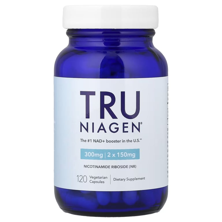 NR,
NR, 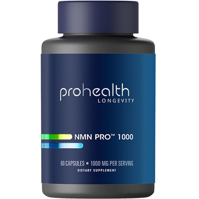 NMN),
NMN), 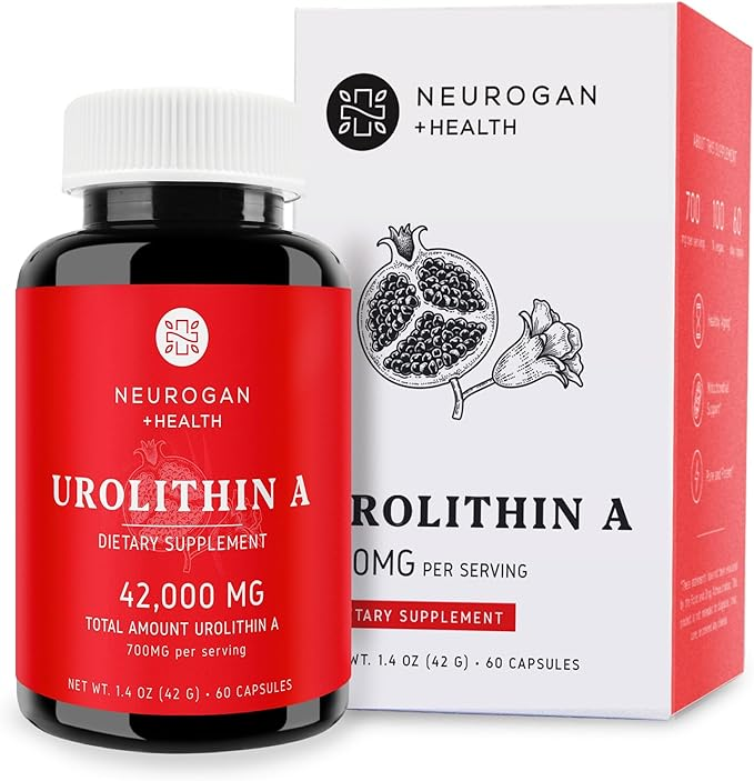 Urolithin A,
 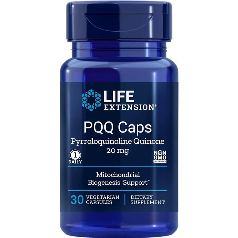 PQQ , and
  MOTS-c.
Mitochondrial biogenesis is one of the few aging mechanisms you can directly counteract. The cellular machinery for building new mitochondria stays functional throughout life, it just needs the signal. Without that signal, mitochondrial mass slowly declines. With it, you can keep mitochondrial density at levels comparable to people decades younger.
Mitophagy
mitophagy (mito = mitochondria, phagy = eating), is the process of cleaning out old mitochondria and it's the cell's quality-control system for mitochondria specifically.
When a mitochondrion accumulates enough damage that it's leaking electrons, producing excess free radicals, and generating less ATP than it consumes, the cell tags it for removal. The damaged mitochondrion gets engulfed by a membrane, broken down inside a lysosome, and its components get recycled into raw materials for building new ones. A new, functional mitochondrion can then take its place through biogenesis.
Without functioning mitophagy, damaged mitochondria accumulate. They keep leaking electrons, keep producing free radicals, keep dragging down ATP output per cell. The cell ends up with more mitochondria by count but fewer that actually work properly, and the dysfunctional ones poison the environment for the healthy ones.
Mitophagy also slows down with age.
What stimulates mitophagy:
- Fasting. The strongest known dietary trigger. Extended fasting (16+ hours) activates AMPK and inhibits mTOR, both of which switch the cell into cleanup mode
- Exercise. Particularly endurance training. The cellular stress of sustained energy demand activates the same pathways
- Caloric restriction. Sustained mild caloric deficit produces the same signal as fasting on a smaller scale
- Urolithin A. A compound produced when gut bacteria metabolise ellagic acid (from pomegranates, walnuts, raspberries). The most direct mitophagy-stimulating compound with reasonable human evidence, and the one with the strongest data of the targeted interventions
- Spermidine. A compound found in wheat germ and aged cheese, also stimulates mitophagy through related pathways
Mitochondrial decline with age
Mitochondrial dysfunction is one of the most consistent findings in aging research.
- Total mitochondrial mass drops as biogenesis signals weaken
- The remaining mitochondria become less efficient at producing ATP, generating more free radicals
- Mitochondrial DNA damage accumulates, partly because mtDNA sits next to where free radicals are generated.
- Mitophagy slows down, so damaged mitochondria stick around longer.
- ATP output per cell falls as a result of all of the above. By age 70 in untrained populations, ATP production is roughly half what it was at 20
Subjectively this shows up as the things people associate with "getting older": fatigue, reduced exercise tolerance, slower recovery, brain fog, reduced metabolic rate, and the general sense that you have less energy than you used to. The cellular reality behind that experience is your mitochondria producing less ATP per cell than they used to.
The same decline is the mechanism behind several major diseases:
- Neurodegeneration. Brain neurons are extremely energy-hungry and fail when mitochondria fail.
- Type 2 diabetes. Mitochondrial dysfunction in muscle and liver impairs glucose uptake and drives insulin resistance.
- Heart failure. Cardiac muscle never stops contracting and is utterly dependent on continuous mitochondrial ATP output.
- Sarcopenia. The age-related loss of muscle mass is partly driven by declining mitochondrial function in muscle fibres.
The good news is this is one of the most modifiable parts of aging. Consistent endurance training, resistance training, periods of caloric restriction or fasting, cold exposure, adequate sleep, and avoiding the things that accelerate mitochondrial damage (chronic alcohol, processed food, sedentary lifestyle, chronic stress, smoking) can preserve mitochondrial function into your 60s and 70s at levels comparable to untrained 30-40 year-olds.
Most longevity-focused interventions work by either preserving existing mitochondrial function or driving the production of new mitochondria.
mTOR vs AMPK
Cells are constantly choosing build new things, or conserve resources and clean up and it's made by two opposing signalling pathways called mTOR and AMPK that read the cell's energy status and switch its activity accordingly.
These two switches govern almost everything cells do. Whether you build muscle after training. Whether you store fat or burn it. Whether you grow or repair. How long you live. mTOR and AMPK are the load-bearing decisions that determine all of it.
mTOR
mTOR (mechanistic target of rapamycin) is the cell's "build" switch. When mTOR is active, the cell ramps up protein synthesis, builds more ribosomes, and increases its overall manufacturing capacity. When mTOR is suppressed, the cell stops building and shifts into maintenance and recycling mode.
mTOR gets activated by signals:
- Amino acids, particularly leucine. This is why protein intake (and leucine specifically) is one of the strongest direct activators of muscle protein synthesis.
- Insulin and IGF-1. Both signal "energy is available, build" and turn mTOR on.
- Mechanical tension on cells. Resistance training mechanically stresses muscle cells, which activates mTOR through a separate pathway.
- Anabolic hormones. Testosterone and growth hormone both ultimately funnel into mTOR activation in muscle tissue.
mTOR also plays a role in cell growth, division, and survival across nearly every tissue and it's why people who eat plenty of protein and train regularly build muscle. It's also one of the reasons chronic overactivation (constant feeding, constant amino acid availability, constant high insulin) accelerates aging, the cell never gets the off-signal it needs to switch into repair and cleanup.
AMPK
AMPK (AMP-activated protein kinase) is mTOR's opposite. If mTOR is the "build" switch, AMPK is the "conserve and burn" switch.
AMPK reads the cell's energy status by tracking how much ATP has been spent. Every time the cell uses ATP for anything, the molecule loses a phosphate group and becomes AMP (adenosine monophosphate). The ratio of AMP to ATP is essentially the ratio of spent currency to available currency, which tells the cell how much of its energy reserves are still in hand.
AMP doesn't stay AMP. The cell keeps recycling: AMP gets recharged back to ADP, ADP back to ATP, mostly through ATP synthase in the mitochondria. The pool of ATP/ADP/AMP is constantly cycling, and you cycle through roughly your body weight in ATP every day.
When AMPK turns on, the cell:
- Increases glucose uptake (pulls more glucose out of the bloodstream into the cell for fuel, independent of insulin)
- Increases fat burning (breaks down stored fat for energy)
- Reduces liver glucose production (the body stops manufacturing new glucose because the cell is in conservation mode)
- Slows fat and cholesterol synthesis (no new storage when resources are tight)
- Triggers autophagy and mitophagy (the cell cleans up damaged components and recycles them)
AMPK essentially activates in response to anything that drops cellular energy: fasting, caloric restriction, sustained exercise, cold exposure. It's also the primary target of  Metformin and
  Berberine , two of the most widely-used insulin-sensitising compounds. Both work by inhibiting mitochondrial complex I (mildly reducing ATP production), raising the AMP:ATP ratio, and forcing AMPK to behave as if fuel is scarce. This is why these compounds improve insulin sensitivity and lower blood sugar even though they're not insulin, they're triggering the same glucose uptake and fat burning that exercise does, through the same cellular sensor.
mTOR and AMPK directly inhibit each other. Activating one suppresses the other. This is the seesaw at the centre of metabolic biology:
- Eating, especially carbs and protein, raises insulin and amino acids → mTOR up, AMPK down → cell is in build and storage mode
- Fasting, sustained exercise, caloric restriction → AMP rises → AMPK up, mTOR down → cell is in burn and cleanup mode
You can't be in both states at the same time. The cell is always biased toward one or the other, and most metabolic interventions are ultimately moving the seesaw in one direction.
And why fasting genuinely changes how cells behave (sustained AMPK activation drives cleanup and fat burning). Why "constant snacking" is metabolically problematic (mTOR never gets to switch off) and why prolonged endurance training improves insulin sensitivity (sustained AMPK activation rewires glucose handling).
The healthiest pattern long-term is oscillation, periods of mTOR activation (training, eating, growing) alternated with periods of AMPK activation (fasting, fasted training, sleep, caloric variation). Chronic activation of either one alone is problematic. Chronic mTOR drives aging through insufficient cleanup. Chronic AMPK depletes building capacity and impairs muscle maintenance. The cells need both states, just not at the same time, and not stuck in one indefinitely.
Cellular Communication
Cells don't operate in isolation. They constantly receive signals from outside (hormones, nutrients, mechanical forces, neighbouring cells) and send signals out. The mTOR/AMPK section covered the two big internal switches that respond to those signals but there are many more:
IGF-1
IGF-1 (insulin-like growth factor 1) is a growth signal produced primarily by the liver in response to growth hormone, but also produced locally inside tissues like muscle, bone, and brain. Local IGF-1 production within a tissue acts as a direct growth signal for the cells in that tissue.
IGF-1 activates mTOR (the build switch covered above), which is why it's such a powerful driver of muscle growth, tissue repair, and cell proliferation. It also increases glucose uptake into cells, suppresses programmed cell death (apoptosis), and stimulates the production of new muscle cells from satellite cells.
Testosterone increases local IGF-1 production within muscle tissue, which is one of the ways it amplifies muscle growth beyond its direct effects on gene expression.
Apoptosis
Apoptosis is programmed cell death, the body's controlled mechanism for removing damaged, infected, or unnecessary cells. Unlike necrosis (uncontrolled cell death from injury), apoptosis is clean and orderly. The cell dismantles itself from the inside in a regulated sequence, and is quietly absorbed by neighbouring cells without triggering inflammation.
Apoptosis is essential for normal tissue maintenance, development, and preventing damaged cells from becoming cancerous. Roughly 50-70 billion cells die through apoptosis every day in an adult body, and get replaced through normal cell division. This is how the body keeps tissues fresh, removes cells that aren't working properly, and prevents the accumulation of damaged or pre-cancerous cells.
Apoptosis can be triggered from inside the cell (when DNA damage is detected, or when the cell becomes dysfunctional) or from outside (when immune cells detect a problem). The decision to trigger apoptosis is one of the most important quality-control mechanisms in the body, when it fails to fire when it should, damaged cells accumulate and can become cancerous. When it fires too aggressively, healthy cells get killed off prematurely (which is part of what happens in some neurodegenerative diseases).
Growth signals like IGF-1, insulin, and several anabolic hormones suppress apoptosis in their target tissues, keeping cells alive longer under stress. This is one of the mechanisms behind why training and adequate hormonal status protect muscle and brain tissue with age.
Glucose uptake
Glucose uptake is how cells acquire their primary fuel. Glucose circulating in the blood can't enter most cells freely, it needs transporter proteins (primarily GLUT4 in muscle and fat cells) to carry it across the cell membrane.
Insulin is the main signal that tells cells to move GLUT4 transporters to the cell surface and start absorbing glucose. IGF-1 does something similar through a related pathway. AMPK activates GLUT4 directly without needing insulin, which is one reason exercise pulls glucose into muscle even when insulin is low.
When cells become insulin resistant, they stop responding properly to the insulin signal. GLUT4 stays inside the cell instead of moving to the surface, glucose stays in the blood (blood sugar rises), and the cells themselves get starved of fuel despite glucose being abundant. This is the core mechanism of type 2 diabetes and is directly linked to testosterone, body composition, and chronic inflammation.
Methylation
Methylation is the chemical transfer of a methyl group (CH3, one carbon plus three hydrogen atoms) from one molecule onto another. It's tiny, just one carbon and three hydrogens, but attaching that single piece to the right spot transforms what the receiving molecule does.
Methylation works in four main ways across the body:
- It activates molecules. Adrenaline is just noradrenaline with one methyl group stuck on. The body converts one neurotransmitter into the next by methylating it. Same logic builds melatonin from serotonin.
- It silences genes. Adding methyl groups to specific stretches of DNA silences those genes, removing them switches them back on. This is DNA methylation, and it's one of the main mechanisms behind epigenetics and biological aging. The "epigenetic clocks" that estimate biological age measure the methylation pattern at a few hundred specific DNA sites that predictably change with age, some gain methyl tags over time, others lose them. The pattern produces a number that correlates with chronological age, and the gap between that estimated age and your actual age is itself predictive of mortality and chronic disease risk.
- It marks molecules for clearance. Oestrogen and other hormones get tagged with methyl groups in the liver, which packages them into forms the body can safely excrete. Poor methylation means hormones don't get cleared properly and accumulates
- It completes the construction of structural molecules like creatine, phosphatidylcholine, and CoQ10.
The body runs millions of methylation reactions per second across every cell.
Cell Damage
Cells get damaged constantly. This is normal. The body has repair systems for everything, the question is whether damage is staying within what those systems can handle or accumulating faster than they can keep up.
Oxidative stress
Your body produces free radicals constantly. The electron transport chain leaks a small percentage of electrons that react with oxygen to form superoxide, the most common free radical. Other sources include immune activity, environmental toxins, UV radiation, and certain enzymatic reactions. This is normal physiology. The defence system is built to handle it.
The problem starts when production outpaces neutralisation. That imbalance is called oxidative stress, and it's considered one of the primary drivers of aging.
Under normal conditions, antioxidant enzymes neutralise free radicals as they're produced, and there's no net damage. Under oxidative stress, the excess radicals attack cell membranes, proteins, and DNA, and damage accumulates faster than the body can repair it.
What tips the balance toward oxidative stress:
- UV radiation. One of the biggest external triggers, which is why sun exposure ages skin so aggressively
- Chronic inflammation. Immune activity generates free radicals as a byproduct, so the two systems amplify each other (covered in the next section)
- Poor diet. Low in the nutrients that fuel antioxidant enzymes (copper, zinc, selenium)
- Pollution and environmental toxins. External sources of free radical generation
- Chronic psychological stress. Cortisol elevation increases free radical production
- Mitochondrial dysfunction. Damaged mitochondria leak more electrons, producing more free radicals, in a self-reinforcing cycle
- Aging itself. The defence system gets weaker while production stays the same or rises
Intense exercise also generates a burst of free radicals. Paradoxically, this is partly why exercise is good for you. Moderate, transient oxidative stress from training forces the body to upregulate its antioxidant defence system, making it stronger over time. It's only chronic, unmanaged oxidative stress that causes damage.
Antioxidants
The antioxidant defence system is the body's mechanism for managing internal chemical damage, specifically the toxic byproducts of energy production. Unlike the immune system which fights external threats, this system is purely internal housekeeping.
The body produces three primary antioxidant enzymes to neutralise free radicals inside cells:
- SOD (superoxide dismutase), the first line of defence. Neutralises superoxide, the most common free radical. Requires copper and zinc to function
- Catalase, breaks down hydrogen peroxide (the byproduct of SOD's work) into plain water
- Glutathione peroxidase, protects cell membranes from oxidative damage. Requires Selenium to function
These enzymes do most of the work. Dietary antioxidants (vitamin C, vitamin E, polyphenols from vegetables, berries, and olive oil) act as supporting players, they're external free radical neutralisers that work alongside the enzymatic system but don't replace it. This is also why supplementing high-dose antioxidants doesn't reliably extend lifespan in studies, the enzymatic system is doing the load-bearing work, and dietary antioxidants add to it but don't substitute for it.
The cell also continuously repairs damaged DNA and replaces damaged cells through normal turnover, which is the other side of the equation.
Chronic inflammation
Inflammation isn't inherently bad. In the short term it's one of the body's most important defence mechanisms. When the immune system detects damage or infection, it triggers a controlled inflammatory response to isolate the threat, recruit immune cells, and begin repair. The problem is when that response never fully turns off.
Cytokines are signalling molecules cells use to communicate. In the context of inflammation, the key ones are TNF-α (tumour necrosis factor alpha) and IL-6 (interleukin-6). Think of them as chemical text messages between cells: "come here," "start attacking," "stop attacking," "start repairing."
Evolutionarily, cytokines were designed to spike hard and fast in response to acute threats (infection, wound, predator attack), then drop back to baseline once the threat was resolved. The system was built for on/off cycles, not continuous low-level activation. Modern life keeps the signal running.
TNF-α is one of the master switches of inflammation. Despite the name, it's not just about tumours. It's one of the first alarm signals released when the immune system detects damage. It tells other immune cells to come to the site, triggers swelling, and activates other inflammatory pathways. Very useful acutely. Very destructive when chronically elevated. Chronic TNF-α elevation:
- Accelerates aging across every tissue (the same fibroblast suppression and collagen breakdown that damages skin happens in joints, blood vessels, and organs)
- Directly interferes with insulin signalling (which is why chronic inflammation and metabolic dysfunction are tightly linked)
- Promotes inflammation in blood vessel walls, contributing to atherosclerosis
- Crosses the blood-brain barrier and promotes neuroinflammation, impairing memory, focus, and mood
- Suppresses muscle protein synthesis and promotes breakdown, which is why chronically inflamed people lose muscle even without trying
IL-6 amplifies the immune response and tells the liver to produce acute-phase proteins, including fibrinogen, a clotting factor that affects blood viscosity and cardiovascular risk. Chronically elevated IL-6 raises fibrinogen, which have shown to be a stronger predictor of cardiovascular death than LDL cholesterol.
What keeps inflammation chronically elevated in modern life:
- Poor sleep (even one bad night measurably elevates IL-6 and TNF-α)
- Chronic psychological stress
- Processed food and seed oils (drive low-grade gut inflammation that feeds systemic cytokine elevation)
- Visceral fat (fat tissue around organs continuously secretes IL-6 and TNF-α)
- UV-damaged skin cells send continuous inflammatory signals
- Lack of movement impairs the body's ability to resolve inflammation
- Smoking
- Aging itself (cytokine levels trend upward with age, a phenomenon called "inflammaging")
Given modern lifestyle factors (processed food, poor sleep, sedentary work, chronic stress, visceral fat), a significant portion of the population runs chronically elevated TNF-α and IL-6.
What it feels like day to day: tiredness despite enough sleep, brain fog, joints that feel stiff in the morning, skin that ages faster, getting sick frequently, low mood and reduced motivation, slower recovery from training. None of this shows up on a standard checkup. It only shows up on a blood test measuring CRP (C-reactive protein), IL-6, or fibrinogen.
DNA damage
DNA damage is the downstream consequence of everything above. Oxidative stress generates free radicals that attack DNA directly. Chronic inflammation suppresses the repair systems that would normally fix that damage. And the consequence is DNA instructions getting corrupted.
What causes DNA damage:
- Free radicals. Steal electrons from DNA bases, distorting their shape and breaking the base pairing. The most common source of damage
- UV radiation. UV photons fuse adjacent thymine bases together, creating a structural deformity called a thymine dimer that the reading machinery can't process properly
- Replication errors. Every time a cell divides, it copies 3 billion base pairs. Even with extraordinary accuracy, the process occasionally inserts the wrong base
- Chemical mutagens. Cigarette smoke, alcohol metabolites, and certain industrial chemicals react directly with DNA bases and alter their chemistry
The body repairs roughly 10,000 to 1,000,000 DNA lesions per cell per day. The repair mechanisms are extraordinarily efficient under normal conditions, which is why most people don't get cancer despite constant DNA damage. Three primary repair systems run continuously, scanning the genome:
- Base excision repair, cuts out single damaged bases and replaces them
- Nucleotide excision repair, removes larger sections of damaged DNA including UV-induced thymine dimers
- Mismatch repair, catches replication errors where the wrong base was inserted during copying
What determines how well the repair system works: age (efficiency declines significantly), micronutrients (repair enzymes require zinc, magnesium, and folate to function), sleep (the majority of DNA repair happens during deep sleep), and oxidative stress and chronic inflammation (high free radical load overwhelms repair capacity, and inflammation directly suppresses repair gene expression).
When the repair system can't keep up, one of three things happens when a cell tries to read a damaged section:
- Misread instruction. The ribosome reads a damaged base and inserts the wrong amino acid into the protein being built. The protein comes out the wrong shape and either malfunctions or doesn't work at all. If this happens in a critical enzyme, the downstream consequences compound
- Stalled machinery. Some damage is severe enough that the reading machinery stops. The gene can't be expressed, the protein doesn't get made. If that protein was essential, the cell either stops functioning or triggers apoptosis
- Permanent mutation. The most dangerous outcome. If a damaged section gets copied before it can be repaired, the damage gets baked into the copy permanently. Every cell that descends from that cell carries the corrupted instruction
A single mutation is usually harmless, the body has redundancy built in. But mutations accumulate over a lifetime. When enough accumulate in the genes that control cell division (specifically, the genes that tell cells when to stop dividing), a cell can lose that stop signal entirely and start dividing uncontrollably. That's cancer.
This is why cancer risk rises so dramatically with age. It's not age itself, it's the accumulated mutation load from decades of damage gradually overwhelming the repair system. Lifestyle is either accelerating or slowing that accumulation.
Cellular senescence
Below the threshold of cancer, accumulated DNA damage still matters. Cells with enough damage stop dividing permanently, entering a state called senescence. This sounds protective, and in the short term it is. A senescent cell can't become cancerous because it can't divide.
The problem is that senescent cells don't die cleanly. They stay alive and secrete a continuous cocktail of inflammatory signals into surrounding tissue, including TNF-α, IL-6, and enzymes that break down structural proteins. This is called the senescence-associated secretory phenotype (SASP). The senescent cells damage neighbouring cells and can push them toward senescence too. It's contagious in slow motion.
Senescent cells accumulating over time are one of the primary drivers of aging:
- Accelerated tissue aging. Senescent cells in skin secrete inflammatory signals locally, suppressing nearby fibroblasts and breaking down collagen. Skin thins, loses firmness, heals slower. Same process in joints, blood vessels, and organs
- Organ decline. Senescent cells accumulating in liver, kidneys, lungs, and cardiovascular tissue progressively impair function. Organs still work but less efficiently and less resiliently
- Chronic inflammation amplification. SASP feeds the chronic inflammation cycle, which feeds further DNA damage, which produces more senescent cells. Another self-reinforcing loop
The four forms of damage in this section all share most of the same triggers: poor sleep, chronic stress, processed food, UV exposure, sedentary lifestyle, alcohol, smoking, and the gradual decline of repair systems with age.
Improving any of them tends to improve the others, because they're not separate problems. They're four angles on the same underlying issue, cellular damage outpacing the body's capacity to fix it.
Fibrosis
Fibrosis is one of the most common forms of long-term tissue damage and the underlying mechanism behind a huge fraction of chronic disease. When tissue is damaged, fibroblasts arrive and lay down collagen to repair it, this is normal. In fibrosis, the signal to stop never comes. Fibroblasts keep producing collagen indefinitely, replacing functional tissue with stiff, disorganised scar tissue. Once enough has been laid down, the tissue can no longer do its original job.
Fibrosis shows up across the body:
- Liver cirrhosis. Repeated damage from alcohol, fatty liver disease, or hepatitis triggers continuous fibroblast activity. Functional liver tissue gets replaced with scar tissue, the organ progressively loses its ability to detoxify, produce proteins, and process nutrients
- Lung fibrosis. The lung's elastic tissue gets replaced by stiff scar tissue, losing the elasticity needed to exchange oxygen. This is what drives COPD progression and idiopathic pulmonary fibrosis
- Kidney fibrosis. Functional filtering tissue gets replaced, eventually leading to chronic kidney disease
- Cardiac fibrosis. Scar tissue accumulating in heart muscle reduces its ability to contract efficiently, a major driver of heart failure
- Vascular fibrosis. Stiffening of artery walls from accumulated scar tissue contributes to hypertension and atherosclerosis
- Skin scarring. The most visible form, but the same underlying mechanism
The core problem is always the same: chronic inflammation keeps TGF-β (transforming growth factor beta) running continuously. TGF-β is the primary signalling molecule that tells fibroblasts to produce collagen and keep producing it. In a normal acute injury, TGF-β spikes and drops.
The body has its own brake on this process: a protein called decorin, produced by fibroblasts as part of normal tissue maintenance. Decorin binds directly to TGF-β and neutralises it, physically grabbing the molecule and preventing it from signalling more collagen production. In chronic disease states, TGF-β is being produced faster than decorin can neutralise it, the brake exists but the signal is too strong. Chronic inflammation makes this worse by directly suppressing decorin production. IL-6 and TNF-α both downregulate the decorin gene, which is why chronic inflammation leads so directly to fibrosis. The inflammation simultaneously drives the fibrotic signal and weakens the body's main mechanism for stopping it.
Fibrosis is one of the main reasons chronic disease tends to be irreversible.
Stem cells
A stem cell is a cell that hasn't fully committed to a specific identity yet. Most cells in your body have a fixed job, a skin cell will always be a skin cell, a muscle cell will always be a muscle cell. Stem cells are different. They're held in reserve, dormant, waiting for the signal to become whatever the body needs.
Stem cells have two properties no other cell has:
Self-renewal. When a stem cell divides, it can produce more stem cells indefinitely. Most cells divide a limited number of times and eventually die. Stem cells can keep dividing and replenishing the stem cell pool without that limit.
Potency. A stem cell can become different cell types depending on what signals it receives. A skin stem cell can become various skin cell types. A muscle stem cell can become a muscle cell. Some stem cells (the most powerful ones) can become almost any cell type in the body.
Stem cells sit on a spectrum based on how many cell types they can become:
- Totipotent. Can become any cell in the body including the placenta. Only the fertilised egg and its very first divisions. Extremely short window
- Pluripotent. Can become almost any cell type in the body. Embryonic stem cells. This is where most of the early stem cell therapy hype came from
- Multipotent. Can become several related cell types but not any cell. Most adult stem cells fall here. Bone marrow stem cells produce all blood cell types, skin basal cells produce all skin layer cells
- Unipotent. Can only produce one cell type but can still self-renew. Muscle satellite cells are the classic example
Most of the stem cells in your body right now are multipotent adult stem cells. They sit quietly in specific locations called niches, activating only when tissue needs repair.
How stem cells work in the body
Stem cells are a tiny minority of cells in any tissue. They sit dormant in their niches most of the time. When tissue is damaged or normal cell turnover requires replacement, signals reach the stem cell niche and activate the cells.
When a stem cell activates, it typically divides asymmetrically: one daughter cell stays behind as a stem cell (preserving the pool), and the other differentiates into the needed cell type. This way the body gets new functional cells without depleting the stem cell reserve.
Your body replaces approximately 3.8 million cells every second to maintain itself. Most of this turnover runs on the back of stem cell activity, even though stem cells themselves are rare. They're the engine behind continuous tissue renewal across the entire body.
Satellite cells
The clearest practical example of stem cells in action is in muscle tissue. Satellite cells are unipotent stem cells that sit on the outside of skeletal muscle fibres, dormant until activated by mechanical stress (training) or hormonal signals (testosterone, IGF-1).
When activated, a satellite cell divides. One copy stays behind to replenish the satellite cell pool. The other fuses with the existing muscle fibre, donating its nucleus to the larger cell. Muscle fibres need lots of nuclei to drive local protein synthesis, and adding more nuclei is one of the main ways muscles grow.
The extra nuclei persist even after the stimulus that caused them is removed. This is the cellular basis of muscle memory, the well-documented phenomenon where previously trained muscle regains size faster than untrained muscle. The nuclei from previous training are still there waiting to be reactivated.
The satellite cell pool is finite and declines with age, which is one of the main reasons older adults struggle to build and maintain muscle even with the same training stimulus.
Stem cells and aging
Stem cells age too, and this is one of the most important hallmarks of biological aging. Three things happen:
The stem cell pool shrinks. The body has fewer stem cells available with age, both because they get used and because they don't replicate as readily as they did in youth. Less reserve means less repair capacity.
The stem cells that remain become less responsive. They sit in their niches but respond more sluggishly to activation signals. Even when damage occurs, the repair response is slower and less complete than it would have been at a younger age.
The niche deteriorates. The local environment that supports stem cells, the surrounding cells, signalling molecules, and structural matrix, gets degraded by chronic inflammation, fibrosis, and oxidative damage. Even healthy stem cells can't function properly in a damaged niche.
The combination is why aging tissue heals slower, regenerates less completely, and accumulates damage faster. It's not just that more damage is happening, it's that the cells responsible for fixing damage have themselves been worn down.
Stem cell therapy
The theory behind stem cell therapy is pluripotent stem cells could theoretically replace any damaged tissue. Heart cells lost after a heart attack. Dopamine neurons lost in Parkinson's. Insulin-producing cells in diabetes.
Turning that theoretical promise into safe, reliable clinical treatments has proven enormously difficult. The cells need to go to the right place, differentiate into exactly the right cell type, integrate properly with existing tissue, and not become cancerous. Most early clinical applications produced modest results or failed outright.
What's actually working clinically right now is bone marrow stem cell transplants for blood cancers (the original stem cell therapy, decades old), limited applications for corneal repair, some emerging work in cartilage and skin regeneration.
The interventions that genuinely affect stem cell function long-term are the same ones that affect most of the rest of cellular biology: avoiding chronic inflammation, controlling oxidative stress, sleeping well, training consistently, eating adequately. They respond to the same environment as the rest of your cells.
 Made with Bullet