Bloodwork is how you objectively track what's happening inside your body. Lab reference ranges are population averages that include unhealthy people, so "normal" on a lab report doesn't necessarily mean optimal. The functional optimal ranges listed here are narrower and represent where you actually want to be for performance, health, and longevity. Always test fasted in the morning for the most consistent and accurate results unless otherwise noted.
Hormones
Total Testosterone
First-line blood test for assessing androgen status. Total testosterone measures all testosterone in the blood: free, albumin-bound, and SHBG-bound combined. It's the starting point, but it doesn't tell the full story on its own because the majority of circulating testosterone is bound to proteins and unavailable to enter cells.
Optimal range (men): 500-900 ng/dL. The lab reference range typically starts at 264-300 ng/dL, but this lower end includes unhealthy, obese, and elderly men. A 30-year-old man at 310 ng/dL is technically "normal" by lab standards but is functionally low. Where you feel best is individual, but most men report optimal energy, libido, and body composition in the 500-900 range.
Optimal range (women): 15-70 ng/dL (premenopausal). Declines gradually from the late twenties onward and drops roughly 50% by menopause.
What low means: Fatigue, low libido, poor recovery from training, brain fog, loss of muscle mass, increased body fat (particularly visceral), depressed mood, irritability. In women, low testosterone presents as low libido, flat mood, reduced energy, and loss of muscle tone.
What high means: truly elevated total testosterone is rare and usually indicates a testicular tumour or adrenal issue.
When to test: Fasted, in the morning (before 10am). Testosterone follows a circadian rhythm and peaks in the early morning, afternoon testing can read 20-30% lower.
Free Testosterone
The testosterone actually available to enter cells and bind to androgen receptors. Only about 2-4% of total testosterone circulates unbound (free). This is the fraction that's doing the work, driving muscle growth, libido, mood, and all the effects described on the hormonal system page. You can have a total testosterone of 600 ng/dL and still feel terrible if most of it is locked up by SHBG.
Optimal range (men): There's no universally agreed functional optimal range because assays vary, but generally 15-25 pg/mL by equilibrium dialysis (the gold standard method) or 50-150 pg/mL by direct immunoassay (the more common but less accurate method). The numbers vary significantly between labs and methods, so always compare against the specific lab's reference range and track trends over time rather than fixating on a single number.
Optimal range (women): Typically 0.5-5 pg/mL by equilibrium dialysis. The Free Androgen Index (FAI), which is the ratio of total testosterone to SHBG, is the preferred clinical measure for women because absolute free testosterone levels are so low that standard assays struggle with precision.
What low means: All the symptoms of low testosterone (fatigue, low libido, poor recovery, brain fog, loss of muscle, mood issues) can appear even with "normal" total testosterone if free testosterone is low. This is the most common scenario that gets missed: a man walks into a clinic, total T comes back at 500 ng/dL, the doctor says "you're fine," but his SHBG is sky-high and his free T is in the gutter.
What high means: free testosterone rises with total testosterone and can be pushed high. There's no established upper limit for clinical harm from free testosterone alone, but very high levels mean more substrate available for conversion to DHT and oestradiol, so downstream markers need to be monitored.
When to test: Same as total testosterone, fasted, morning. Always test alongside total testosterone and SHBG to see the full picture. If your lab offers equilibrium dialysis, use it. If only direct immunoassay is available, it's still useful for tracking trends but the absolute numbers are less reliable.
SHBG (Sex Hormone Binding Globulin)
A protein produced by the liver that binds testosterone, oestradiol, and DHT in the bloodstream. SHBG-bound hormones are functionally unavailable, they can't enter cells or bind to receptors. SHBG acts as a reservoir and controlled-release system, preventing hormones from being cleared too quickly while only allowing a small percentage to be free at any given time.
Optimal range (men): 20-50 nmol/L. Within this range, SHBG is doing its job as a buffer without excessively locking up testosterone. Below 20 often comes packaged with insulin resistance and metabolic dysfunction. Above 50 can leave free testosterone low even when total testosterone looks adequate.
Optimal range (women): 40-120 nmol/L. Women naturally have higher SHBG because oestrogen stimulates its production. Very high SHBG in women (common on oral contraceptives) can suppress free testosterone enough to cause low libido and flat mood.
What high SHBG means: Less free testosterone and oestradiol available. Common causes include elevated oestrogen (oral contraceptives, liver disease), hyperthyroidism, aging, low caloric intake, and low body fat. A man with total testosterone of 600 ng/dL and SHBG of 70 nmol/L may have less bioavailable testosterone than a man at 450 ng/dL with SHBG of 25 nmol/L.
What low SHBG means: More free testosterone technically, but also faster metabolic clearance and it usually indicates underlying metabolic problems. Common causes include insulin resistance, obesity, type 2 diabetes and hypothyroidism. Low SHBG in the context of metabolic dysfunction is not a good sign even though it means higher free testosterone on paper.
When to test: Fasted, morning, alongside total and free testosterone. SHBG is the missing piece that explains why total testosterone alone can be misleading. If total T looks normal but symptoms persist, SHBG tells you whether that testosterone is actually available.
Important context: SHBG doesn't just bind testosterone. It also binds oestradiol and DHT, though with different affinities. DHT binds most strongly, then testosterone, then oestradiol. This means high SHBG preferentially locks up androgens over oestrogen, which can shift the effective androgen-to-oestrogen ratio even when the absolute numbers look balanced.
Bioavailable Testosterone
Free testosterone plus albumin-bound testosterone combined. Albumin binds testosterone loosely, and as blood flows through capillary beds in tissues, the testosterone dissociates from albumin fairly easily and can enter cells. So bioavailable testosterone represents the total fraction that can actually get into tissues and do work, roughly two-thirds of total testosterone.
Why it matters: Bioavailable testosterone is a more complete picture than free testosterone alone. Free T only captures the 2-4% that's completely unbound. Bioavailable T captures both the free fraction and the albumin-bound fraction that's readily available in tissues. Some clinicians prefer bioavailable testosterone as the primary functional marker because it better reflects what's actually reaching androgen receptors.
Optimal range (men): Roughly 130-680 ng/dL, though this varies significantly by assay and lab. Like free testosterone, track trends over time rather than fixating on a single number.
How it's calculated: Most labs don't measure bioavailable testosterone directly. It's calculated from total testosterone, SHBG, and albumin levels using a formula. This is another reason to always test total T, free T, and SHBG together, the calculation depends on all three.
Oestradiol (E2)
The primary and most potent form of oestrogen. In men, oestradiol is produced by aromatase converting testosterone, primarily in fat tissue, brain, bone, and testes. In women, the ovaries are the primary source before menopause. Oestradiol is critical for bone density, cardiovascular health, brain function, joint health, libido, collagen production, and lipid metabolism in both sexes.
Optimal range (men): 20-35 pg/mL is where most men feel best, though it's highly individual. Some function well slightly outside this range. The ratio of testosterone to oestradiol matters as much as the absolute number.
Optimal range (women): Fluctuates across the menstrual cycle. Early follicular phase: 30-50 pg/mL. Pre-ovulatory peak: 200-400 pg/mL. Mid-luteal phase: 100-300 pg/mL. Postmenopausal: 5-25 pg/mL. Interpreting a woman's oestradiol result requires knowing where she is in her cycle.
What low means (men): Joint pain and stiffness, dry skin, flat mood and emotional numbness, low libido, fatigue, poor cognitive function, accelerated bone loss.
What high means (men): Water retention and bloating, sensitive or puffy nipples (early gynecomastia), mood swings and emotional volatility, reduced libido, increased anxiety, fat gain particularly around the chest and hips.
What low means (women): Menopausal symptoms (hot flashes, night sweats, vaginal dryness), accelerated bone loss, increased cardiovascular risk, skin thinning, joint stiffness, cognitive changes, mood disruption.
When to test: Fasted, morning.
Important context: If oestradiol is elevated, the first approach should be optimising body composition (less body fat means less aromatase).
DHT (Dihydrotestosterone)
Produced when 5-alpha reductase converts testosterone to DHT, primarily in the prostate, skin, hair follicles, and liver. DHT is 3-5 times more potent than testosterone at the androgen receptor. It contributes to libido, drive, confidence, and physique hardness, but it also drives hair loss in genetically susceptible individuals, prostate growth, and acne. DHT acts primarily as a local hormone in the tissues where it's produced rather than circulating systemically like testosterone.
Optimal range (men): 30-80 ng/dL. Some men need the higher end for optimal libido and drive, others feel fine at the lower end. Individual variation is significant.
What low means: Low libido despite adequate free testosterone and oestradiol, lack of assertiveness and drive, softer physique, poor erectile quality, thinning body hair. Low DHT is most common in men taking 5-alpha reductase inhibitors (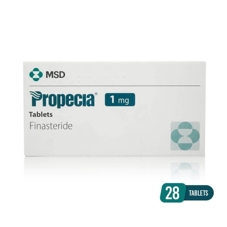 Finasteride,
 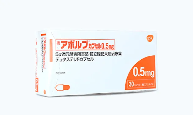 Dutasteride) for hair loss or BPH.
What high means: Accelerated hair loss (in genetically susceptible individuals, not universally), oily skin, acne, prostate enlargement with urinary symptoms (frequent urination, weak stream), increased irritability.
When to test: Fasted, morning, alongside total and free testosterone. DHT is not included in standard hormone panels and needs to be specifically requested. It's most useful to test when libido is low despite adequate testosterone and oestradiol (to check if DHT is unexpectedly low), when experiencing hair loss or prostate symptoms (to confirm DHT is elevated), or when taking finasteride/dutasteride (to verify the drug is actually reducing DHT as intended).
Important context: DHT's role in adult libido is debated. Men with congenital 5-alpha reductase deficiency appear to have normal sexual function, but men who lose DHT pharmacologically through finasteride report sexual side effects at rates of 2-15% in clinical trials (with placebo groups reporting 1-7%, making the nocebo effect a genuine confounding factor). The balance between testosterone, DHT, and oestradiol matters more than any single number.
Prolactin
A peptide hormone produced by lactotroph cells in the anterior pituitary. Its primary biological function is stimulating milk production (also the reason some bodybuilders can at times squeeze milk from their tits). Elevated prolactin is one of the most common and underdiagnosed causes of low libido, sexual dysfunction, and emotional flatness in men, even when testosterone and oestrogen look normal.
Prolactin and dopamine have an inverse relationship. Dopamine actively suppresses prolactin release. When dopamine tone drops, prolactin rises because the brake is removed. The symptoms (low libido, flatness, no motivation) are largely dopamine-deficit symptoms. Prolactin also independently suppresses GnRH, which reduces LH and therefore testosterone production.
Optimal range (men): 2-18 ng/mL. Ideally in the single digits.
Optimal range (women): 2-29 ng/mL. Higher baseline reflects oestrogen's stimulatory effect on lactotroph cells.
What high means: Low libido and sexual dysfunction even with adequate testosterone and oestradiol, erectile dysfunction, emotional flatness, lack of motivation, and secondary HPG suppression (prolactin directly suppresses GnRH, lowering LH and testosterone). Levels above 25-30 ng/mL in men warrant investigation. Levels above 100 ng/mL strongly suggest a prolactinoma (a benign pituitary tumour that produces prolactin autonomously).
What low means: Not typically a clinical concern.
Common causes of elevation: Medications that block D2 dopamine receptors (antipsychotics are the most common), SSRIs (serotonin stimulates prolactin release), opioids, stress, elevated oestrogen (oestrogen stimulates lactotroph cell proliferation, increases prolactin gene transcription, and reduces D2 receptor expression making cells less responsive to dopamine), prolactinomas, and 19-nortestosterone derivatives (nandrolone, trenbolone) which raise prolactin through progesterone receptor activation on lactotroph cells.
When to test: Fasted, morning. Can be transiently elevated by stress, sleep, food, and nipple stimulation, so repeat an elevated result to confirm.
Cortisol
The body's primary stress hormone, produced in the adrenal cortex in response to ACTH from the pituitary. Cortisol follows a circadian rhythm: it peaks in the early morning (the cortisol awakening response that helps you wake up) and declines throughout the day, reaching its lowest point around midnight. Disrupted cortisol patterns, elevated at night or flattened throughout the day, are markers of HPA axis dysregulation.
Optimal range: Morning cortisol (drawn before 10am): 10-20 mcg/dL. The absolute number matters less than the pattern, ideally high in the morning, low in the evening. A morning cortisol below 5 mcg/dL suggests adrenal insufficiency. A morning cortisol above 25 mcg/dL suggests excessive HPA axis activation or Cushing's.
What high means: Chronically elevated cortisol promotes muscle breakdown, visceral fat accumulation, insulin resistance, HPG axis suppression (reduced testosterone), immune suppression, hippocampal damage (memory and cognition), bone loss, collagen degradation, impaired sleep, and compromised recovery from training. Chronic stress, overtraining, sleep deprivation, and excessive caloric restriction all keep cortisol elevated.
What low means: Persistently low cortisol can indicate adrenal insufficiency (primary or secondary), HPA axis burnout from prolonged chronic stress, or medication effects (abrupt withdrawal from long-term corticosteroid use). Symptoms include severe fatigue, dizziness, low blood pressure, brain fog, and inability to handle stress.
When to test: Fasted, first thing in the morning (before 10am). Cortisol is highly time-sensitive because of the circadian rhythm, an afternoon sample will read much lower and isn't comparable to a morning draw. For a fuller picture, a four-point salivary cortisol test (morning, noon, afternoon, evening) maps the daily rhythm and reveals patterns that a single blood draw misses.
Important context: Always interpret cortisol alongside DHEA-S. The cortisol-to-DHEA-S ratio tells you more about the stress-hormone balance than either number alone. High cortisol with low DHEA-S suggests the adrenals are being driven hard but the precursor hormone pool is depleting, a pattern common in chronic stress, overtraining, and aging.
DHEA-S
The most abundant circulating steroid hormone in the body, produced primarily in the zona reticularis of the adrenal cortex. DHEA-S serves as a precursor that peripheral tissues convert into testosterone and oestrogen. It's also a marker of adrenal function and the stress-hormone balance. In women, adrenal DHEA-S is the source of roughly 50% of total testosterone production through peripheral conversion, making it particularly important for female androgen status.
DHEA-S and cortisol are both produced by the adrenal cortex, both driven by ACTH. In acute stress, both rise together. In chronic, sustained stress, their trajectories diverge: cortisol tends to stay elevated while DHEA-S declines over time, creating the imbalanced ratio often described as "adrenal fatigue."
Optimal range (men): 280-640 mcg/dL, though this declines with age. DHEA-S follows the steepest age-related decline of any hormone, dropping roughly 2-3% per year from its peak in the mid-twenties. By 70-80, levels can be 10-20% of peak.
Optimal range (women): 65-380 mcg/dL, also declining with age.
What low means: Low DHEA-S with normal or high cortisol suggests the adrenals are being driven hard but the precursor pool is depleting, meaning less raw material is available for downstream conversion to sex hormones. This pattern is common in chronic stress, overtraining, and aging. Low DHEA-S is also independently associated with increased cardiovascular risk, reduced immune function, and cognitive decline.
What high means: Elevated DHEA-S can indicate adrenal overproduction (congenital adrenal hyperplasia, adrenal tumours) or exogenous DHEA supplementation. In women, elevated DHEA-S is one of the markers checked when investigating PCOS or other androgen-excess conditions.
When to test: Fasted, morning. DHEA-S is relatively stable throughout the day (unlike cortisol), so timing is less critical, but morning fasted is still the most consistent. Always interpret alongside cortisol to assess the raI gutio.
Important context: A low DHEA-S is one of the reasons people with chronic stress can have hormonal symptoms that don't fully resolve by addressing testosterone alone. The chronic stress pattern that caused the low DHEA-S is still doing damage through cortisol's effects on sleep, immune function, gut health, insulin resistance, and recovery.
Progesterone
Typically thought of as a female reproductive hormone, but it has relevant functions in both sexes. In women, progesterone is produced by the corpus luteum after ovulation and rises during the luteal phase. It's essential for maintaining the uterine lining for potential pregnancy and is a precursor to allopregnanolone, a neurosteroid that enhances GABAergic signalling in the brain, producing calming, anxiolytic, and sleep-promoting effects. In men, small amounts are produced by the testes and adrenal glands. Progesterone also converts to neurosteroid metabolites in men, contributing to sleep quality and anxiety regulation.
Optimal range (men): 0.3-1.2 ng/mL. Not routinely tested unless specific symptoms suggest it may be relevant.
Optimal range (women): Varies dramatically across the cycle. Follicular phase: <1 ng/mL. Mid-luteal phase (roughly 7 days after ovulation): 5-20 ng/mL. A mid-luteal progesterone below 3 ng/mL suggests anovulation (no egg was released that cycle). Postmenopausal: <0.5 ng/mL.
What low means (women): Anovulatory cycles, luteal phase defects (short or inadequate second half of the cycle), difficulty maintaining early pregnancy, premenstrual anxiety and insomnia (the GABA-enhancing neurosteroid signal is insufficient), irregular cycles.
What low means (men): Poor sleep quality, increased anxiety, and mood instability that doesn't fully resolve with adequate testosterone and oestradiol. Low progesterone in men is rarely tested for but may be relevant in men on finasteride, which blocks 5-alpha reductase and therefore also blocks the conversion of progesterone to its calming neurosteroid metabolites.
What high means (men): Elevated progesterone in men can indicate adrenal overproduction or exogenous use. Some clinicians use low-dose progesterone for its anti-oestrogenic properties and sleep-promoting effects, though this is not standard practice.
When to test (women): Mid-luteal phase (day 21 of a 28-day cycle, or 7 days after confirmed ovulation). Testing at the wrong point in the cycle will give a meaninglessly low result.
When to test (men): Only when symptoms suggest it may be relevant (unexplained anxiety, insomnia, or mood issues not explained by other markers). Fasted, morning.
Metabolic
Fasting Glucose
The amount of glucose in your blood after at least 8-12 hours without eating. Glucose is the body's primary fuel source, and the pancreas regulates blood sugar through insulin (lowers glucose by telling cells to absorb it) and glucagon (raises glucose by telling the liver to release it). Fasting glucose tells you how well this system is maintaining baseline blood sugar when no food is coming in.
Optimal range: 70-90 mg/dL. Lab reference ranges typically go up to 100 mg/dL before flagging "pre-diabetic," but functional optimal is tighter. Consistently sitting at 95-99 mg/dL means the system is already under strain even if the lab says "normal."
What high means: Fasting glucose above 100 mg/dL indicates impaired fasting glucose (pre-diabetes). Above 126 mg/dL on two separate tests is diagnostic for type 2 diabetes. Elevated fasting glucose means either the pancreas isn't producing enough insulin, or the cells have become resistant to insulin's signal and the liver is releasing glucose it shouldn't be. Chronic hyperglycaemia is directly toxic to the endothelium (blood vessel lining), accelerates atherosclerosis, damages nerves, and drives the formation of advanced glycation end-products (AGEs) that cross-link collagen and stiffen arteries.
What low means: Below 70 mg/dL is hypoglycaemia. Symptoms include shakiness, sweating, confusion, irritability, and in severe cases loss of consciousness. Reactive hypoglycaemia (blood sugar crashing a few hours after eating, particularly after high-carb meals) can indicate early insulin dysregulation where the pancreas is overproducing insulin in response to glucose spikes.
When to test: Fasted for at least 8 hours, morning. Fasting glucose alone is a blunt instrument. It only tells you where blood sugar sits at baseline, not how the system handles a glucose load or how hard insulin is working to maintain that number. Always interpret alongside fasting insulin and HbA1c for the full picture.
Fasting Insulin
The amount of insulin the pancreas is producing to maintain your fasting glucose level. This is the marker most people never test, and it's arguably more important than fasting glucose for catching metabolic problems early.
Here's why: glucose can stay "normal" for years while insulin is climbing. The pancreas compensates for developing insulin resistance by producing more and more insulin to force the same amount of glucose into cells. Fasting glucose only rises once the pancreas can no longer keep up. By the time fasting glucose is flagged as pre-diabetic, insulin resistance has been building for years. Fasting insulin catches the problem much earlier.
Optimal range: 2-8 mIU/L. Lab reference ranges often go up to 25 mIU/L before flagging it, which is far too generous. A fasting insulin of 15 with a normal fasting glucose means the pancreas is already working overtime to maintain that glucose level. The system is under strain.
What high means: Insulin resistance. The cells (muscle, liver, fat) are becoming less responsive to insulin's signal, so the pancreas produces more to compensate. High fasting insulin is associated with increased visceral fat storage, elevated triglycerides, suppressed SHBG (and therefore less bioavailable testosterone), increased inflammation, elevated blood pressure, and accelerated cardiovascular risk. It's one of the earliest and most sensitive markers of metabolic dysfunction.
What low means: Normal insulin sensitivity (good), or in rare cases inadequate insulin production (type 1 diabetes or late-stage type 2 where the pancreas has burned out from years of overproduction).
When to test: Fasted for at least 8 hours, morning. Most standard metabolic panels don't include fasting insulin, you need to request it specifically. Combine with fasting glucose to calculate HOMA-IR.
Important context: If you only test one metabolic marker beyond basic bloodwork, make it fasting insulin. It reveals the trajectory of your metabolic health years before fasting glucose or HbA1c start moving.
HbA1c
A measure of average blood sugar over the past 2-3 months. Glucose in the bloodstream sticks to haemoglobin on red blood cells (a process called glycation), and since red blood cells live about 90-120 days, the percentage of haemoglobin that's been glycated reflects how high blood sugar has been running over that lifespan. Where fasting glucose gives you a single snapshot, HbA1c gives you the long-term trend.
Optimal range: 4.0-5.3%. Lab reference ranges consider below 5.7% normal, 5.7-6.4% pre-diabetic, and 6.5%+ diabetic. But functional optimal is lower. Sitting at 5.5-5.6% is technically "normal" but already indicates blood sugar is running higher than it should be.
What high means: Blood sugar has been chronically elevated over the past 2-3 months. This means either insulin isn't being produced adequately, or cells are resistant to insulin's signal and glucose is spending too long in the bloodstream. Chronically elevated blood sugar drives glycation of proteins throughout the body, not just haemoglobin. The same process damages collagen in blood vessel walls (contributing to arterial stiffness through AGE cross-linking), damages nerves (diabetic neuropathy), damages the kidneys (diabetic nephropathy), and damages the retina (diabetic retinopathy). Every 1% increase in HbA1c is associated with significantly increased cardiovascular risk.
What low means: Below 4.0% can indicate hypoglycaemia, haemolytic anaemia (red blood cells being destroyed faster than normal, so there's less time for glycation), or significant blood loss. Unusually low HbA1c in someone not trying to lower it warrants investigation.
When to test: No fasting required, can be drawn at any time of day. This is one of its advantages over fasting glucose. Test every 3-6 months for monitoring, more frequently if actively managing blood sugar or making significant dietary changes.
Important context: HbA1c can be misleading in certain situations. Anything that changes red blood cell lifespan affects the result. Iron deficiency anaemia (smaller, longer-lived red blood cells) can falsely elevate HbA1c. Recent blood loss or haemolytic conditions can falsely lower it. Haemoglobin variants (common in certain ethnic populations) can interfere with some assay methods. If HbA1c doesn't match the clinical picture (symptoms, fasting glucose, fasting insulin), consider these confounders.
HOMA-IR
A calculated score that estimates insulin resistance. You take your fasting glucose and fasting insulin, plug them into a formula (fasting insulin × fasting glucose ÷ 405, when glucose is in mg/dL), and get a single number that tells you how hard your pancreas is working to maintain your blood sugar.
Optimal range: Below 1.0 is excellent insulin sensitivity. 1.0-1.5 is good. 1.5-2.0 is early insulin resistance developing. Above 2.0 is meaningful insulin resistance. Above 3.0 is significant and strongly associated with metabolic syndrome and pre-diabetes.
Why it matters: HOMA-IR catches metabolic dysfunction earlier than fasting glucose or HbA1c because it factors in how much insulin the pancreas is producing to maintain that glucose level. A fasting glucose of 88 mg/dL with a fasting insulin of 4 mIU/L gives a HOMA-IR of 0.87, excellent. A fasting glucose of 92 mg/dL with a fasting insulin of 14 mIU/L gives a HOMA-IR of 3.19, significant insulin resistance, even though both fasting glucose numbers look "normal" on a lab report.
What high means: The cells are becoming resistant to insulin's signal. The pancreas is compensating by producing more insulin to force glucose into cells. This state drives visceral fat accumulation, suppresses SHBG (reducing bioavailable testosterone), increases triglycerides, raises blood pressure, promotes systemic inflammation, and accelerates cardiovascular risk. Insulin resistance is the metabolic foundation under most of the body composition and hormonal problems discussed across this site.
When to calculate: Any time you have fasting glucose and fasting insulin from the same blood draw. Most labs don't calculate it for you, but the formula is simple enough to do yourself.
Lipids
for a deep-dive into how lipids work read about Lipids in our vascular system
Total Cholesterol
The sum of all cholesterol in your blood: LDL, HDL, VLDL, and other lipoprotein fractions combined. Cholesterol itself is essential, your body uses it to build cell membranes, produce steroid hormones (testosterone, oestrogen, cortisol, and every other steroid hormone discussed on this site), synthesise vitamin D, and make bile acids for digesting fat. The problem is never "having cholesterol," it's having the wrong distribution of it.
Optimal range: Below 200 mg/dL is the standard guideline. But total cholesterol alone is a poor predictor of cardiovascular risk because it doesn't tell you the breakdown. A total cholesterol of 220 with high HDL and low LDL is a very different picture than 200 with low HDL and high LDL. Total cholesterol is the headline number, but the story is in the components.
What high means: Could mean elevated LDL (increased atherogenic risk), elevated HDL (protective, not a concern), or both. The number alone doesn't tell you which, so a full lipid panel is always needed. Chronically elevated total cholesterol driven by high LDL is associated with accelerated atherosclerosis.
What low means: Below 150 mg/dL can indicate malnutrition, liver dysfunction, hyperthyroidism, or malabsorption. Very low cholesterol can theoretically impair steroid hormone production since cholesterol is the raw material, though the body is efficient at synthesising its own cholesterol and dietary cholesterol is a smaller contributor than most people think.
When to test: Fasted for at least 8 hours. Part of a standard lipid panel alongside LDL, HDL, and triglycerides. Test every 6-12 months as a baseline, more frequently if managing lipid-related risk or on compounds that affect the lipid profile.
Important context: Total cholesterol is becoming less relevant as a standalone marker. ApoB (which measures the actual number of atherogenic particles) is a far better predictor of cardiovascular risk. If your panel includes ApoB, that tells you more than total cholesterol does.
LDL
LDL (low-density lipoprotein) delivers cholesterol from the liver to cells throughout the body. This is a legitimate and necessary function, cells need cholesterol for membranes, hormone production, and repair. The problem starts when LDL levels are chronically elevated and particles spend too long circulating in the bloodstream.
When there's more LDL than the body needs, particles begin penetrating the walls of blood vessels, particularly in areas where the endothelium is already damaged or inflamed. Once inside the vessel wall, LDL oxidises. The immune system recognises oxidised LDL as a threat and sends macrophages to engulf it. These macrophages become overloaded and turn into foam cells. Foam cells accumulate into fatty streaks, which over years develop into plaques that narrow the artery and can rupture, triggering a clot. This is atherosclerosis, and it's the underlying cause of most heart attacks and strokes.
Optimal range: Below 100 mg/dL for most people. Below 70 mg/dL for anyone with existing cardiovascular risk factors or a family history of heart disease. The lower the lifetime exposure to LDL, the lower the atherosclerotic risk.
What high means: Increased atherogenic particle burden, accelerated plaque formation, higher cardiovascular risk. High LDL can be driven by genetics (familial hypercholesterolaemia), diet (high saturated fat intake), insulin resistance, hypothyroidism, or compounds that affect hepatic lipid metabolism.
What low means: Generally favourable from a cardiovascular perspective. Very low LDL (below 40 mg/dL) is occasionally seen with hyperthyroidism, malabsorption, or liver disease but is rarely a concern in practice.
When to test: Fasted for at least 8 hours, as part of a full lipid panel. Every 6-12 months at baseline, more frequently if actively managing lipids or on compounds that affect the lipid profile.
Important context: Standard LDL on most lab panels is calculated (using the Friedewald equation), not directly measured. This calculation becomes inaccurate when triglycerides are high (above 400 mg/dL) or very low. Direct LDL measurement is available but less common. More importantly, ApoB is a better marker of cardiovascular risk than LDL-C because it counts the actual number of atherogenic particles rather than the cholesterol content within them. Two people with the same LDL-C can have very different particle counts, and it's the particle count that drives plaque formation.
HDL
HDL (high-density lipoprotein) is the reverse transport system. Where LDL delivers cholesterol from the liver out to cells, HDL picks up excess cholesterol from cells and arterial walls and carries it back to the liver to be broken down and excreted. This process is called reverse cholesterol transport, and it's one of the body's primary defences against atherosclerotic plaque buildup.
Optimal range: Above 40 mg/dL is the minimum for men, above 50 mg/dL for women. Functionally, above 50-60 mg/dL is where you want to be. Higher is generally better, though extremely high HDL (above 100 mg/dL) doesn't necessarily provide additional protection and in rare cases may reflect dysfunctional HDL particles that aren't actually performing reverse cholesterol transport effectively.
What high means: Generally protective. Higher HDL is associated with lower cardiovascular risk. Regular aerobic exercise, moderate alcohol consumption, and adequate oestrogen all raise HDL. High HDL is one of the reasons premenopausal women have lower cardiovascular risk than age-matched men.
What low means: Reduced capacity to clear cholesterol from arterial walls, increased cardiovascular risk. Low HDL is one of the defining features of metabolic syndrome alongside high triglycerides, insulin resistance, visceral obesity, and elevated blood pressure. Common causes of low HDL include sedentary lifestyle, smoking, insulin resistance, obesity, very low-fat diets, and supraphysiological androgens.
When to test: Fasted for at least 8 hours, as part of a full lipid panel. Every 6-12 months at baseline, more frequently if on compounds that affect lipids.
Important context: HDL is useful but it's not the whole picture. The ratio of triglycerides to HDL is one of the better surrogate markers of insulin resistance. A triglyceride-to-HDL ratio below 1.0 suggests good insulin sensitivity. Above 2.0 suggests developing insulin resistance. Above 3.0 is strongly associated with metabolic dysfunction.
Triglycerides
Fats circulating in your bloodstream. When you eat more calories than your body needs immediately, the excess is converted to triglycerides, packaged into VLDL particles in the liver, and released into the bloodstream for transport to fat cells for storage. Triglycerides are also released from fat stores between meals to provide energy. The fasting level reflects how much fat the liver is packaging into each VLDL and releasing when no food is coming in.
Optimal range: Below 100 mg/dL. Lab reference ranges consider below 150 mg/dL normal, but functional optimal is lower. Below 80 mg/dL is excellent. Above 150 is elevated, above 200 is high, above 500 is severe and carries risk of pancreatitis.
What high means: The liver is producing and releasing more VLDL (triglyceride-rich particles) than the body is clearing. High triglycerides are driven by excess caloric intake (particularly refined carbohydrates and sugar, which the liver converts to fat very efficiently), insulin resistance (insulin normally suppresses hepatic VLDL production, so when cells are resistant to insulin, the liver keeps packaging and releasing triglycerides), alcohol consumption, obesity, and poorly managed diabetes. High triglycerides are also associated with elevated small dense LDL particles (the most atherogenic subtype), low HDL, and increased cardiovascular risk. Supraphysiological androgen use can also elevate triglycerides depending on the compounds, dose, and diet.
What low means: Good metabolic health, efficient fat clearance. Very low triglycerides (below 40 mg/dL) are occasionally seen with malnutrition, malabsorption, or hyperthyroidism but are rarely a concern.
When to test: Fasted for at least 8-12 hours. Triglycerides are the most food-sensitive marker on a lipid panel, a non-fasted sample can read significantly higher and isn't useful for comparison. Part of a standard lipid panel.
Important context: The triglyceride-to-HDL ratio is one of the most practical surrogate markers of insulin resistance and metabolic health. Below 1.0 is excellent. 1.0-2.0 is acceptable. Above 2.0 suggests developing insulin resistance. Above 3.0 is strongly associated with metabolic syndrome. This ratio is often more informative than either number alone.
ApoB
Apolipoprotein B is a protein found on the surface of every atherogenic lipoprotein particle: LDL, VLDL, IDL, and lipoprotein(a). Each of these particles carries exactly one ApoB molecule, so measuring ApoB directly counts the total number of atherogenic particles in your bloodstream. This is why ApoB is a better predictor of cardiovascular risk than LDL-C (which measures the cholesterol content inside LDL particles, not the number of particles themselves).
Two people can have identical LDL-C readings but very different particle counts. One person might have fewer, larger, cholesterol-rich LDL particles. The other might have many more small, dense LDL particles that each carry less cholesterol but collectively represent a much higher atherogenic burden. Standard LDL-C would read the same for both. ApoB would correctly identify the second person as higher risk because they have more particles capable of penetrating the arterial wall.
Optimal range: Below 90 mg/dL for most people. Below 80 mg/dL for anyone actively optimising cardiovascular health. Below 60 mg/dL for anyone with existing cardiovascular risk factors or family history of heart disease. Some longevity-focused clinicians aim for below 60 mg/dL in everyone regardless of risk profile, on the basis that lifetime atherogenic particle exposure is the primary driver of atherosclerosis.
What high means: More atherogenic particles circulating, greater chance of particles penetrating the arterial wall and initiating plaque formation. Elevated ApoB is driven by the same factors as elevated LDL: genetics, diet, insulin resistance, hypothyroidism, and compounds that affect hepatic lipid metabolism.
What low means: Lower atherogenic particle burden, lower cardiovascular risk. Very low ApoB is not a clinical concern.
When to test: Fasted, as part of an advanced lipid panel. Not included on standard lipid panels at most labs, you need to request it specifically. If your lab only offers a standard lipid panel (total cholesterol, LDL-C, HDL, triglycerides), the non-HDL cholesterol calculation (total cholesterol minus HDL) is a reasonable surrogate for ApoB, though less precise.
Important context: If you can only add one marker to a standard lipid panel, make it ApoB. It's the single best predictor of atherosclerotic cardiovascular risk from a lipid perspective. The shift in cardiology toward ApoB over LDL-C as the primary target reflects the growing understanding that it's the number of particles entering the arterial wall that drives disease, not the amount of cholesterol they carry.
Lp(a)
Lipoprotein(a) is a variant of LDL with an additional protein called apolipoprotein(a) attached to the ApoB on its surface. It's atherogenic for the same reasons LDL is, it carries cholesterol into the artery wall, but worse, it’s harder to clear and competes with plasminogen for binding sites, so it both promotes plaque formation and interferes with the body's natural clot-dissolving mechanisms.
Lp(a) unique is that it's almost entirely genetically determined. Your Lp(a) level is set by your genes, it doesn't respond meaningfully to diet, exercise, or most medications. For now, your level is largely what you were born with.
Optimal range: Below 30 mg/dL (or below 75 nmol/L). Above 50 mg/dL (or 125 nmol/L) is considered elevated and meaningfully increases cardiovascular risk. The units and thresholds vary by assay, so check which unit your lab uses.
What high means: A genetically fixed, lifelong cardiovascular risk factor. Elevated Lp(a) contributes to plaque formation through the same mechanism as LDL (particle penetration into the intima, oxidation, foam cell formation) and additionally promotes clot stability by competing with plasminogen. This combination of pro-atherogenic and pro-thrombotic effects makes elevated Lp(a) particularly dangerous.
What low means: One fewer cardiovascular risk factor to think about.
When to test: Once in a lifetime. Since Lp(a) doesn't change, a single test tells you whether it's a factor in your risk profile or not. It's not included in standard lipid panels, you need to request it specifically.
Important context: Roughly 20% of the global population has Lp(a) levels high enough to meaningfully increase cardiovascular risk, and most don't know it because it's never tested. Lp(a) can explain why some people with otherwise clean lipid panels and healthy lifestyles develop cardiovascular disease. If you have a family history of early heart disease and your standard lipid markers look reasonable, Lp(a) is worth checking. If it's elevated, it shifts the risk management conversation toward more aggressive control of the factors you can modify (ApoB, blood pressure, inflammation, body composition) to offset the genetic burden you can't.
Liver
ALT (Alanine Aminotransferase)
An enzyme found primarily in liver cells. When liver cells are damaged or inflamed, ALT leaks into the bloodstream, so elevated ALT is one of the most sensitive markers of liver stress or damage. It's more liver-specific than AST (which is also found in heart and muscle tissue), making it the first marker most clinicians look at for liver health.
Optimal range: 10-30 U/L. Lab reference ranges often go up to 40-55 U/L, but emerging research suggests the upper "normal" limit should be lower. Consistently sitting at 35-45 U/L with a "normal" flag on the lab report may still indicate subclinical liver stress, particularly from fatty liver disease, alcohol, or hepatotoxic compounds.
What high means: Liver cells are being damaged. Common causes include non-alcoholic fatty liver disease (NAFLD, the most common cause of elevated ALT in the general population, driven by insulin resistance and visceral fat), alcohol consumption, hepatotoxic medications (statins, paracetamol/acetaminophen at high doses, NSAIDs with chronic use), oral anabolic steroids (17-alpha-alkylated compounds like oxandrolone, stanozolol, and dianabol are particularly hepatotoxic because they pass through the liver directly), viral hepatitis, and autoimmune liver conditions. Mildly elevated ALT (30-60 U/L) is common and often reflects fatty liver or mild hepatic stress. Significantly elevated ALT (above 100 U/L) warrants urgent investigation.
What low means: Normal liver cell turnover. Very low ALT (below 7-10 U/L) can occasionally indicate vitamin B6 deficiency (ALT requires B6 as a cofactor) or advanced liver disease where so few functional liver cells remain that there's nothing left to leak.
When to test: Fasted, as part of a standard liver panel (also called hepatic function panel or LFTs). Every 6-12 months at baseline. More frequently if on oral compounds, taking hepatotoxic medications, or consuming alcohol regularly. For anyone running oral anabolics, liver enzymes should be checked before, during, and after the cycle.
Important context: A single elevated ALT reading doesn't mean liver damage is permanent. The liver is one of the most regenerative organs in the body. ALT often normalises once the offending cause is removed (stopping the compound, reducing alcohol, improving metabolic health). Persistent elevation over months is what warrants concern. Always interpret ALT alongside AST, GGT, albumin, and bilirubin for the full picture.
AST (Aspartate Aminotransferase)
Another enzyme involved in amino acid metabolism, but unlike ALT, AST is found in multiple tissues: the liver, heart, skeletal muscle, kidneys, and red blood cells. This makes AST less liver-specific than ALT. An elevated AST can mean liver damage, but it can also mean muscle damage from intense training, a cardiac event, or haemolysis (red blood cell destruction).
Optimal range: 10-30 U/L. Similar to ALT, lab reference ranges often go up to 40 U/L but functional optimal is lower.
What high means: Depends on context. If both ALT and AST are elevated together, it's likely liver-related (fatty liver, alcohol, hepatotoxic compounds). If AST is elevated but ALT is normal or only mildly elevated, it's more likely from muscle damage (heavy training, rhabdomyolysis) or cardiac tissue damage. The AST-to-ALT ratio (also called the De Ritis ratio) helps differentiate: a ratio below 1 (ALT higher than AST) suggests liver disease like NAFLD. A ratio above 2 suggests alcoholic liver disease. A ratio above 1 with both values mildly elevated in someone who trains hard may simply reflect muscle turnover.
What low means: Normal cellular turnover. Not typically a clinical concern.
When to test: Fasted, as part of a standard liver panel alongside ALT. Avoid intense training for 48-72 hours before testing if you want an accurate read on liver health specifically, because heavy resistance training can elevate AST (and to a lesser extent ALT) through normal muscle damage that has nothing to do with the liver. If you've trained hard within 48 hours of a blood draw, expect AST to be elevated and don't panic.
Important context: For anyone who trains regularly, a mildly elevated AST with a normal ALT is almost always from muscle tissue, not the liver. This is one of the most common causes of unnecessary concern on bloodwork in the fitness community. If AST is elevated and you want to confirm it's not liver-related, GGT is the tiebreaker, it's liver-specific and won't be elevated from training.
GGT (Gamma-Glutamyl Transferase)
A liver enzyme involved in glutathione metabolism and amino acid transport across cell membranes. GGT is the most sensitive marker of bile duct obstruction and liver stress from alcohol and certain medications. Its value on a bloodwork panel is as the tiebreaker: when ALT or AST are elevated and you need to know whether it's the liver or something else (like muscle damage from training), GGT answers the question because it's liver and bile duct-specific.
Optimal range: 10-30 U/L. Lab reference ranges often go up to 60-70 U/L, but emerging evidence suggests lower is better and elevated GGT even within the "normal" range is associated with increased cardiovascular and metabolic risk.
What high means: Bile duct obstruction or cholestasis (gallstones, bile duct disease), alcohol consumption (GGT is the most sensitive marker of alcohol intake, even moderate drinking can elevate it), hepatotoxic medications, fatty liver disease, pancreatic disease, and chronic inflammation. Elevated GGT is also independently associated with cardiovascular risk, metabolic syndrome, and type 2 diabetes, suggesting it reflects broader oxidative stress and metabolic dysfunction beyond just liver health.
What low means: Normal liver and bile duct function. Not a clinical concern.
When to test: Fasted, as part of a liver panel alongside ALT, AST, albumin, and bilirubin. Particularly valuable if AST is elevated and you need to determine whether it's liver-related or from training. If GGT is normal and AST is elevated with normal ALT, the AST elevation is almost certainly from muscle tissue, not the liver.
Important context: GGT is one of the first markers to rise with alcohol consumption and one of the first to normalise with abstinence. If you're monitoring the impact of alcohol on your liver, GGT is the most responsive indicator. For anyone on oral anabolics, GGT alongside ALT gives a more complete picture of hepatobiliary stress than ALT alone.
Albumin
The most abundant protein in the blood, produced by the liver. Albumin has two main jobs: maintaining oncotic pressure (preventing fluid from leaking out of blood vessels into surrounding tissues) and acting as a general-purpose transport protein for fat-soluble molecules that can't dissolve in blood on their own, including testosterone, fatty acids, thyroid hormones, bilirubin, and many drugs.
Albumin is also a sensitive marker of liver synthetic function and nutritional status. The liver produces roughly 10-15 grams of albumin per day, and this production requires adequate protein intake and functional liver cells. When either is compromised, albumin drops.
Optimal range: 4.0-5.0 g/dL. Lab reference ranges typically start at 3.5 g/dL, but below 4.0 already suggests suboptimal liver function or nutritional status.
What low means: The liver isn't producing enough albumin. Common causes include chronic liver disease (cirrhosis, hepatitis, fatty liver disease where enough functional liver tissue has been lost to impair production), malnutrition or inadequate protein intake, chronic inflammation (inflammatory cytokines suppress albumin synthesis, so chronically elevated CRP often comes with low albumin), kidney disease (nephrotic syndrome causes albumin to leak into urine), and overhydration (dilutional effect). Low albumin causes oedema (fluid retention, puffy ankles, swollen face) because without adequate oncotic pressure, fluid leaks out of the vasculature into tissues. It also means reduced transport capacity for hormones and drugs.
What high means: Almost always dehydration. When blood volume decreases, the concentration of albumin increases. Genuinely elevated albumin production is extremely rare. If albumin is above 5.0 g/dL, you were probably dehydrated when the blood was drawn.
When to test: Fasted, as part of a liver panel or comprehensive metabolic panel. Albumin changes slowly (half-life of about 20 days), so it reflects chronic status rather than acute changes. A single low reading warrants repeat testing to confirm.
Important context: Albumin is the protein that loosely binds testosterone in the bloodstream (the albumin-bound fraction that makes up the "bioavailable" portion of total testosterone). Low albumin means less transport capacity, which can affect hormone distribution. For anyone with unexpectedly low bioavailable testosterone despite reasonable total and free testosterone, checking albumin is worth doing.
Bilirubin
A yellow pigment produced when the body breaks down old red blood cells. Red blood cells live about 90-120 days, and when they're recycled, the haemoglobin inside them is broken down. The haem portion is converted to bilirubin, which travels to the liver, gets conjugated (made water-soluble), and is excreted through bile into the digestive system. This is what gives stool its brown colour and urine its yellow colour.
Bilirubin appears on a liver panel because the liver is responsible for processing and excreting it. If the liver can't keep up (from liver disease, bile duct obstruction, or excessive red blood cell breakdown), bilirubin accumulates in the blood and eventually deposits in the skin and eyes, causing jaundice (yellowing).
Types: Labs often report total bilirubin, and sometimes break it into direct (conjugated, already processed by the liver) and indirect (unconjugated, not yet processed). This distinction helps identify where the problem is: elevated indirect bilirubin suggests the issue is before the liver (excessive red blood cell breakdown or the liver not conjugating efficiently), while elevated direct bilirubin suggests the issue is after the liver (bile duct obstruction preventing excretion).
Optimal range: Total bilirubin 0.2-1.2 mg/dL. Mildly elevated bilirubin (1.2-3.0 mg/dL) without other liver enzyme abnormalities is very commonly caused by Gilbert's syndrome, a benign genetic condition affecting roughly 5-10% of the population where the liver conjugates bilirubin slightly slower than normal. It's harmless and doesn't require treatment.
What high means: Depends on context. If ALT, AST, and GGT are also elevated, it suggests liver disease or bile duct obstruction. If liver enzymes are normal but bilirubin is mildly elevated, it's most likely Gilbert's syndrome. If bilirubin is very high (above 3.0 mg/dL) with visible jaundice, it warrants urgent investigation regardless of other markers. Elevated bilirubin can also indicate haemolytic anaemia (excessive red blood cell destruction overwhelming the liver's processing capacity).
What low means: Normal bilirubin production and clearance. Not a clinical concern.
When to test: As part of a standard liver panel. No special preparation required beyond standard fasting.
Important context: Interestingly, mildly elevated bilirubin (as in Gilbert's syndrome) may actually be protective. Bilirubin is a potent antioxidant, and people with Gilbert's syndrome have been shown in some studies to have lower rates of cardiovascular disease. So a mildly elevated bilirubin with normal liver enzymes is not something to worry about and may even be quietly working in your favour.
Kidney
Creatinine
A waste product generated by the normal breakdown of creatine phosphate in muscle tissue. Your muscles use creatine phosphate as a rapid energy source during short bursts of activity, and creatinine is the metabolic byproduct of that process. It's produced at a fairly constant rate based on your muscle mass, filtered out of the blood by the kidneys, and excreted in urine. Because production is relatively stable and clearance depends on kidney function, creatinine serves as one of the primary markers of how well your kidneys are filtering.
Optimal range: 0.7-1.2 mg/dL for men, 0.5-1.0 mg/dL for women. Muscular individuals and people supplementing creatine will naturally run higher, sometimes up to 1.3-1.5 mg/dL without any kidney problem. This is one of the most commonly misinterpreted markers on bloodwork for people who train hard.
What high means: Either the kidneys aren't clearing creatinine efficiently (reduced kidney function), or production is elevated (high muscle mass, creatine supplementation, recent intense training, high meat consumption). The clinical concern is kidney function. If creatinine is elevated alongside a low eGFR and elevated BUN, that pattern points toward impaired kidney filtration. If creatinine is mildly elevated but eGFR is reasonable and you're a muscular person taking creatine, it's likely a false alarm. Dehydration can also temporarily elevate creatinine by reducing blood flow to the kidneys.
What low means: Low muscle mass, malnutrition, or liver disease (the liver produces creatine, so impaired liver function can reduce the substrate). Not typically a clinical concern in otherwise healthy people.
When to test: Fasted, well-hydrated, as part of a basic metabolic panel or renal panel. If you supplement creatine, mention it to whoever is interpreting your results, otherwise a mildly elevated creatinine can trigger unnecessary concern and follow-up testing. Avoid intense training for 24-48 hours before testing if you want the most accurate kidney-specific reading.
Important context: Creatinine alone is a blunt tool. It's used to calculate eGFR (which adjusts for age, sex, and race to estimate actual kidney filtration rate), and always needs to be interpreted alongside eGFR and BUN for a complete picture. A muscular man on creatine with a creatinine of 1.4 mg/dL and a normal eGFR has healthy kidneys. The same creatinine in a sedentary man not taking creatine warrants further investigation. If you’re taking creatine, evaluate cystatin C for kidney function instead
eGFR (Estimated Glomerular Filtration Rate)
A math formula calculated from your creatinine result combined with your age, sex, and race. Calculated estimate of how much blood your kidneys are filtering per minute. The glomeruli are tiny filtering units in the kidneys (roughly one million per kidney) that filter waste products, excess fluid, and electrolytes out of the blood while keeping proteins and blood cells in. eGFR uses your creatinine level, age, and sex to estimate how efficiently these filters are working.
Optimal range: Above 90 mL/min/1.73m² is normal kidney function. 60-89 is mildly reduced (stage 2 CKD, often asymptomatic and may be normal for older adults). 30-59 is moderately reduced (stage 3, warrants monitoring and investigation). Below 30 is severely reduced (stage 4-5, serious kidney impairment). Below 15 is kidney failure.
What low means: The kidneys aren't filtering blood as efficiently as they should. This can be caused by chronic kidney disease (from diabetes, hypertension, or autoimmune conditions), acute kidney injury (dehydration, certain medications, rhabdomyolysis from extreme training), or simply aging (eGFR naturally declines with age, losing roughly 1 mL/min per year after age 30). NSAIDs (ibuprofen, naproxen) taken chronically can damage the kidneys and lower eGFR. High-dose creatine supplementation and very high protein intake can also lower eGFR, though in these cases it may partly reflect elevated creatinine production rather than true kidney impairment.
What high means: Not a clinical concern. Very high eGFR can occur in early diabetes (hyperfiltration, where the kidneys are working too hard) or during pregnancy, but above-normal eGFR is not something that gets flagged in practice.
When to test: Calculated automatically any time creatinine is tested, most labs report it alongside creatinine on a basic metabolic or renal panel. No separate test required.
Important context: eGFR has the same limitations as creatinine for muscular individuals and creatine users. Because eGFR is calculated from creatinine, anything that elevates creatinine (high muscle mass, creatine supplementation) will make eGFR appear lower than the kidneys' actual filtration rate. If eGFR comes back mildly low (60-80) in someone who is muscular and takes creatine, it may not reflect real kidney impairment. Cystatin C is an alternative marker that isn't affected by muscle mass and can be tested alongside creatinine for a more accurate eGFR calculation in these cases.
Cystatin C
A small protein produced at a steady rate by virtually every nucleated cell in the body. Like creatinine, cystatin C is filtered out of the blood by the kidneys, so the level circulating in blood reflects how well the kidneys are filtering. Unlike creatinine, cystatin C production is independent of muscle mass, diet, and exercise, which makes it the cleaner kidney marker for anyone whose creatinine is artificially elevated by high muscle mass, creatine supplementation, or high protein intake.
Optimal range: 0.5-1.0 mg/L. Lab reference ranges typically go up to 1.0-1.3 mg/L depending on the assay. Values above 1.0 suggest reduced filtration, above 1.3 indicates meaningful kidney dysfunction. Cystatin C rises with age (like creatinine) and can be used to calculate a cystatin C-based eGFR that isn't confounded by muscle mass.
Why it matters: for muscular individuals, athletes, and anyone supplementing creatine, creatinine-based eGFR chronically underestimates actual kidney function. A muscular man on creatine with a creatinine of 1.4 mg/dL might have a creatinine-based eGFR of 60-70 mL/min (flagged as mildly reduced) while his cystatin C-based eGFR comes back at 95+ mL/min (completely normal). The difference isn't a kidney problem, it's the limitation of using a muscle-derived waste product to estimate filtration in someone with a lot of muscle. Cystatin C cuts through this entirely.
What high means: genuine reduced kidney filtration. Because cystatin C isn't affected by muscle mass or creatine, an elevated cystatin C is a more reliable signal of actual kidney dysfunction than elevated creatinine alone. Cystatin C has also been shown in multiple large studies to be a stronger predictor of cardiovascular events and mortality than creatinine, likely because it captures filtration changes earlier and without the muscle mass confounder.
What low means: normal kidney function.
When to test: fasted, as part of a kidney panel. Not included in standard metabolic panels, must be specifically requested. Particularly worth testing if creatinine or eGFR come back borderline in someone who is muscular, on creatine, or eats high protein. Also useful in older adults where muscle mass is declining and creatinine becomes less reliable (an elderly person can have normal creatinine despite reduced kidney function because they have less muscle producing less creatinine).
Important context: when both creatinine-based eGFR and cystatin C-based eGFR are reported, the average of the two is generally considered the most accurate estimate of true filtration rate in healthy adults. For anyone on creatine long-term, cystatin C is the marker that cuts through the creatinine artefact and gives a clean read on actual kidney function. If your doctor is worried about your kidney numbers because of elevated creatinine and you're on creatine, cystatin C is the test that resolves the question.
BUN (Blood Urea Nitrogen)
A measure of urea in the blood. When your body breaks down protein (from food or from muscle tissue being catabolised), the nitrogen-containing amino acids are processed by the liver into urea, which is then filtered out by the kidneys and excreted in urine. BUN reflects both how much protein is being broken down and how well the kidneys are clearing the waste.
Optimal range: 7-20 mg/dL. Some labs report up to 25 mg/dL as normal.
What high means: Either the kidneys aren't clearing urea efficiently (kidney dysfunction), or protein breakdown is elevated. Common causes of elevated BUN include dehydration (the most common and most benign cause, concentrated blood means concentrated urea), high protein diet, GI bleeding (blood in the digestive tract is digested like protein, producing a urea load), kidney disease, heart failure (reduced blood flow to the kidneys), and catabolic states where muscle tissue is being broken down (severe illness, overtraining, high cortisol). Certain medications can also raise BUN.
What low means: Low protein intake, liver disease (the liver can't convert amino acids to urea efficiently), overhydration, or malnutrition. Not typically a concern in otherwise healthy people eating adequate protein.
When to test: Fasted, as part of a basic metabolic panel or renal panel. Always interpret alongside creatinine and eGFR.
Important context: The BUN-to-creatinine ratio helps differentiate the cause of kidney-related elevations. A normal ratio is roughly 10:1 to 20:1. A ratio above 20:1 with elevated BUN suggests a pre-renal cause (dehydration, heart failure, GI bleeding) where the kidneys themselves are fine but the upstream conditions are creating more urea or less blood flow to the kidneys. A ratio within normal range with both BUN and creatinine elevated suggests intrinsic kidney disease. For people who train hard and eat high protein, a mildly elevated BUN with normal creatinine and normal eGFR is usually just reflecting protein turnover and is not a kidney concern.
Blood / Haematology
Haematocrit
The percentage of your blood volume that's occupied by red blood cells. If your haematocrit is 45%, it means 45% of your blood is red blood cells and 55% is plasma and other components. It's one of the most important markers for anyone on exogenous testosterone because testosterone directly stimulates red blood cell production through erythropoietin (EPO).
Optimal range: 40-50% for men, 36-44% for women.
What high means: Blood is thicker and more viscous. Above 50% warrants attention. Above 54% is a significant cardiovascular risk, the thicker blood increases resistance in blood vessels, raises blood pressure, and increases the risk of clots, stroke, and heart attack. High haematocrit is one of the most common and most dangerous side effects of exogenous androgens. It's also caused by dehydration (concentrated blood reads as higher haematocrit), living at high altitude (the body produces more red blood cells to compensate for lower oxygen), sleep apnoea (intermittent oxygen deprivation stimulates EPO), smoking, and polycythaemia vera (a bone marrow disorder).
What low means: Anaemia. Not enough red blood cells to deliver adequate oxygen to tissues. Causes include iron deficiency (the most common cause globally), B12 or folate deficiency, chronic kidney disease (kidneys produce less EPO), chronic inflammation (inflammatory cytokines suppress red blood cell production), blood loss, and bone marrow disorders. Symptoms include fatigue, weakness, pale skin, shortness of breath, and dizziness.
When to test: As part of a complete blood count (CBC). Always test well-hydrated, because dehydration artificially inflates haematocrit and can trigger unnecessary concern.
Important context: If haematocrit is creeping above 50%, the standard interventions are therapeutic phlebotomy (donating blood or a medical blood draw to reduce red blood cell volume), and ensuring adequate hydration. It's one of the few markers where an overlooked elevation can cause an acute cardiovascular event.
Haemoglobin
The iron-containing protein inside red blood cells that actually binds and carries oxygen. Each red blood cell contains roughly 270 million haemoglobin molecules, and each haemoglobin molecule can carry four oxygen molecules. Haemoglobin picks up oxygen in the lungs (where oxygen concentration is high) and releases it in tissues (where oxygen concentration is low). It's the functional counterpart to haematocrit: haematocrit tells you what percentage of your blood is red blood cells, haemoglobin tells you how much oxygen-carrying capacity those cells actually have.
Optimal range: 14-17.5 g/dL for men, 12-15.5 g/dL for women.
What high means: Increased oxygen-carrying capacity, which sounds good but carries the same risks as elevated haematocrit: thicker blood, increased viscosity, higher clot risk. High haemoglobin and high haematocrit move together since haemoglobin is inside the red blood cells that haematocrit is measuring. The same causes apply: dehydration, high altitude, sleep apnoea, smoking, polycythaemia vera.
What low means: Anaemia. The blood can't carry enough oxygen to meet tissue demands. There are many types depending on the cause. Iron deficiency anaemia (most common globally) produces small, pale red blood cells with less haemoglobin per cell. B12 or folate deficiency anaemia produces large, immature red blood cells that don't function properly. Anaemia of chronic disease occurs when chronic inflammation suppresses red blood cell production. Symptoms across all types include fatigue, weakness, pale skin, shortness of breath on exertion, dizziness, and poor exercise tolerance.
When to test: As part of a complete blood count (CBC). Same timing as haematocrit.
Important context: Low haemoglobin with adequate iron, B12, and folate should prompt investigation into chronic inflammation (check CRP), kidney function (kidneys produce EPO), or other underlying causes. In women, heavy menstrual periods are a common and often overlooked cause of low haemoglobin through chronic blood loss depleting iron stores.
Red Blood Cell Count
The total number of red blood cells per volume of blood, typically reported in millions per microlitre (M/uL). Where haematocrit tells you the percentage of blood occupied by red blood cells and haemoglobin tells you oxygen-carrying capacity, the RBC count is the raw cell number. All three are related but can diverge in certain conditions.
Optimal range: 4.5-5.5 M/uL for men, 4.0-5.0 M/uL for women.
What high means: The body is producing more red blood cells than normal. Causes mirror elevated haematocrit: exogenous androgens (EPO stimulation), dehydration, high altitude, sleep apnoea, smoking, polycythaemia vera. A high RBC count with proportionally high haemoglobin and haematocrit is a straightforward picture, more cells, more haemoglobin, thicker blood. A high RBC count with low haemoglobin per cell suggests iron deficiency, the body is producing extra cells to compensate for each cell carrying less oxygen.
What low means: Fewer red blood cells being produced or more being destroyed or lost. Causes include iron, B12, or folate deficiency, chronic kidney disease (reduced EPO), bone marrow disorders, chronic inflammation, autoimmune haemolytic anaemia (the immune system destroying red blood cells), and blood loss. Symptoms are those of anaemia: fatigue, weakness, pale skin, shortness of breath.
When to test: As part of a complete blood count (CBC). Same timing as haematocrit and haemoglobin.
Important context: The RBC count is most useful when interpreted alongside haemoglobin, haematocrit, and red blood cell indices (MCV, MCH, MCHC) which describe the size and haemoglobin content of individual cells. These indices help differentiate types of anaemia. A low RBC count with small cells (low MCV) points toward iron deficiency. A low RBC count with large cells (high MCV) points toward B12 or folate deficiency. A low RBC count with normal-sized cells points toward chronic disease, kidney issues, or bone marrow problems.
White Blood Cell Count
The total number of white blood cells (leukocytes) in your blood, your immune system's circulating workforce. White blood cells detect and respond to infections, damaged tissue, and foreign substances. The count reflects how active or suppressed your immune system is at the time of the draw.
A standard CBC reports the total WBC count and a differential breakdown by cell type. The main types and what they do:
Neutrophils (50-70% of WBCs) are the first responders to bacterial infections. They're the most abundant white blood cell and the ones that surge during acute infections. A high neutrophil count (neutrophilia) usually means an active bacterial infection or acute inflammation. A low count (neutropenia) means increased vulnerability to bacterial infections.
Lymphocytes (20-40%) are the adaptive immune system's main cells, including T-cells (which coordinate immune responses and kill infected cells), B-cells (which produce antibodies), and natural killer cells. Elevated lymphocytes often indicate viral infections. Low lymphocytes can indicate immune suppression from chronic stress, cortisol elevation, HIV, or immunosuppressive drugs.
Monocytes (2-8%) are the cells that become macrophages when they enter tissues, engulfing pathogens and debris and presenting antigens to T-cells to activate the adaptive immune response. Elevated monocytes can indicate chronic infection, chronic inflammation, or autoimmune conditions.
Eosinophils (1-4%) are involved in parasitic infections and allergic responses. Elevated eosinophils suggest allergies, asthma, parasitic infection, or certain autoimmune conditions.
Basophils (<1%) are the rarest white blood cell, involved in allergic and inflammatory responses. Rarely clinically significant on routine bloodwork.
Optimal range (total WBC): 4,500-11,000 cells/uL. Functionally, 5,000-8,000 is a healthy resting range. Consistent counts at the lower or upper end of the reference range deserve attention.
What high means: The immune system is activated. Acute infection (bacterial or viral), chronic inflammation, physical stress, smoking, corticosteroid use (which causes a redistribution of white cells rather than true immune activation), and leukaemia (very high counts, typically above 20,000-30,000 with abnormal cell types). Chronically mildly elevated WBC (9,000-11,000) without obvious infection often reflects low-grade systemic inflammation, which connects to the chronic inflammation section on the cellular biology page.
What low means: Immune suppression. The body has fewer circulating immune cells available to respond to threats. Causes include chronic stress and elevated cortisol, viral infections (including HIV), autoimmune conditions, bone marrow disorders, chemotherapy, and immunosuppressive medications. Testosterone at supraphysiological levels also suppresses white blood cell activity, particularly T-cells and B-cells.
When to test: As part of a complete blood count (CBC). No special preparation required beyond standard fasting. If WBC is abnormal, the differential breakdown (which cell types are elevated or suppressed) is what guides interpretation.
Important context: A single elevated WBC reading during an acute cold or infection is meaningless for health assessment. What matters is the trend when you're healthy. Chronically elevated WBC at rest, without active infection, is an independent marker of cardiovascular risk and systemic inflammation.
Platelets
Cell fragments produced by megakaryocytes in the bone marrow. Platelets are the clotting system's first responders. When a blood vessel is damaged, platelets stick to the exposed tissue, aggregate together, and form a plug. This plug is then reinforced by fibrin (a protein produced from fibrinogen in the clotting cascade) to create a stable clot that stops bleeding. The count reflects how many platelets are circulating and available for this job.
Optimal range: 150,000-400,000 /uL. Most healthy people sit between 200,000-300,000.
What high means (thrombocytosis): More platelets circulating, which can increase clotting risk. Mild elevation (400,000-500,000) is commonly reactive, meaning it's a temporary response to something else: acute infection or inflammation, iron deficiency (the bone marrow overproduces platelets alongside red blood cells when iron is low), recent surgery or trauma, intense exercise, or chronic inflammatory conditions. Persistent or significantly elevated platelet counts (above 500,000-600,000) can indicate a myeloproliferative disorder (the bone marrow is overproducing) and warrants haematological investigation.
What low means (thrombocytopenia): Fewer platelets available for clotting, which increases bleeding risk. Bruising easily, prolonged bleeding from cuts, petechiae (tiny red dots on the skin from capillary bleeding), and in severe cases spontaneous bleeding. Causes include viral infections (which can temporarily suppress platelet production), autoimmune conditions (immune thrombocytopenic purpura, where the body destroys its own platelets), liver disease (the liver produces thrombopoietin which stimulates platelet production, and also clears old platelets, so liver dysfunction disrupts the balance), bone marrow disorders, certain medications (heparin, some antibiotics, chemotherapy), heavy alcohol consumption, and splenic sequestration (an enlarged spleen traps and removes too many platelets from circulation).
When to test: As part of a complete blood count (CBC). No special preparation required.
Important context: For anyone on compounds that stress the liver (oral anabolics, heavy alcohol use), monitoring platelets alongside liver enzymes gives a more complete picture of hepatic health. The liver's role in producing thrombopoietin and clearing platelets means that platelet count can be an indirect signal of liver function that doesn't show up on standard liver enzyme panels.
Ferritin
The body's primary iron storage protein. Ferritin stores iron inside cells (primarily in the liver, spleen, and bone marrow) and releases it as needed for red blood cell production, enzyme function, and oxygen transport. The ferritin level in your blood reflects how much iron you have in reserve, making it the best single marker of iron status.
Optimal range: 30-300 ng/mL for men, 30-200 ng/mL for women. Many functional health practitioners prefer to see ferritin above 50-80 ng/mL for optimal energy and performance. Below 30 is functionally depleted even if haemoglobin hasn't dropped yet. Below 15 is definitive iron deficiency.
What low means: Depleted iron stores. Low ferritin is the earliest stage of iron deficiency, it drops before haemoglobin or red blood cell count are affected. Symptoms include fatigue, poor exercise tolerance, brain fog, hair thinning, restless legs, cold intolerance, and in women, worsening of menstrual symptoms. Causes include inadequate dietary iron (particularly in vegetarians and vegans), chronic blood loss (heavy menstrual periods in women, GI bleeding in men), poor absorption (coeliac disease, low stomach acid, excessive tea or coffee consumption with meals which inhibits iron absorption), intense training (exercise-induced iron loss through sweat, GI microbleeding, and foot-strike haemolysis in runners), and high-dose zinc supplementation without monitoring (zinc competes with iron for absorption).
What high means: Either iron overload or inflammation. Ferritin is an acute phase reactant, meaning it rises during inflammation and infection as part of the immune response, regardless of actual iron stores. A high ferritin with high CRP likely reflects inflammation, not true iron overload. A high ferritin with normal CRP and elevated transferrin saturation suggests genuine iron overload, which can be caused by hereditary haemochromatosis (a genetic condition affecting roughly 1 in 200 people of Northern European descent where the body absorbs too much iron), excessive iron supplementation, or repeated blood transfusions. Iron overload damages the liver, heart, pancreas, and joints because excess iron generates free radicals through the Fenton reaction.
When to test: Fasted, morning. Always interpret alongside a full iron panel (serum iron, TIBC, transferrin saturation) and CRP to distinguish true iron status from inflammatory elevation. A single high ferritin reading during an illness or infection is not meaningful for assessing iron stores.
Important context: Ferritin is one of the most commonly suboptimal markers in women of reproductive age due to monthly menstrual blood loss. A woman presenting with fatigue, brain fog, and hair thinning should have ferritin checked before anything else. Many women are told their iron is "fine" because their haemoglobin is still in range, but their ferritin is sitting at 15-25 ng/mL, which is functionally depleted. For men, unexplained high ferritin with normal inflammatory markers warrants screening for hereditary haemochromatosis, especially in those of Northern European descent.
Thyroid
TSH (Thyroid Stimulating Hormone)
Produced by the anterior pituitary, TSH is the signal that tells the thyroid gland to produce thyroid hormones (T3 and T4). TSH is the first-line screening marker for thyroid function because it responds to thyroid hormone levels through a negative feedback loop: when thyroid hormones are low, TSH rises (the pituitary is shouting louder at the thyroid to produce more). When thyroid hormones are high, TSH drops (the pituitary backs off because there's enough). This makes TSH the most sensitive early indicator of thyroid dysfunction, it moves before the thyroid hormones themselves go out of range.
Optimal range: 0.5-2.5 mIU/L. Lab reference ranges often go up to 4.0-5.0 mIU/L, but functional optimal is tighter. A TSH of 3.5-4.5 with a "normal" flag may already indicate the thyroid is underperforming and the pituitary is compensating. Some people feel significantly better when TSH is brought below 2.0.
What high means: The pituitary is working harder to stimulate the thyroid, which means the thyroid isn't producing enough on its own. This is hypothyroidism (underactive thyroid). Causes include Hashimoto's thyroiditis (an autoimmune condition where the immune system attacks the thyroid, the most common cause), iodine deficiency, medications (lithium, amiodarone), and previous thyroid surgery or radiation. Symptoms: fatigue, weight gain, cold intolerance, constipation, dry skin, hair thinning, brain fog, depression, low libido, slow recovery, and elevated SHBG suppression (hypothyroidism lowers SHBG, increasing free testosterone but in a metabolically dysfunctional context).
What low means: The pituitary is backing off because thyroid hormones are already high. This is hyperthyroidism (overactive thyroid). Causes include Graves' disease (an autoimmune condition that stimulates the thyroid), thyroid nodules producing excess hormone, or excessive thyroid medication. Symptoms: weight loss, anxiety, tremor, heat intolerance, rapid heart rate, insomnia, irritability, and diarrhoea. Very low TSH (below 0.1 mIU/L) with elevated free T3/T4 warrants urgent investigation.
When to test: No fasting required, but morning testing is preferred because TSH has a circadian rhythm (peaks overnight, lowest in the afternoon). If monitoring thyroid medication, test before taking the morning dose.
Important context: TSH alone doesn't tell the full story. A normal TSH with symptoms of hypothyroidism warrants checking free T3 and free T4 to see whether the thyroid hormones themselves are optimal or just scraping by within the reference range. Thyroid function also directly affects testosterone: hypothyroidism lowers SHBG and alters the testosterone-to-oestrogen balance, while hyperthyroidism raises SHBG and can reduce bioavailable testosterone.
Free T3
The active thyroid hormone. The thyroid produces mostly T4 (thyroxine), which is a relatively inactive prohormone. T4 gets converted to T3 (triiodothyronine) in peripheral tissues, primarily the liver, kidneys, and muscle, by enzymes called deiodinases. T3 is roughly 3-5 times more metabolically active than T4. It's the hormone that actually binds to thyroid receptors inside cells and drives metabolic rate, energy production, body temperature regulation, and protein synthesis. Free T3 measures the unbound, active fraction circulating in the blood.
Optimal range: 3.0-4.0 pg/mL. Lab reference ranges typically go from 2.0-4.4 pg/mL, but sitting at 2.2-2.5 with symptoms of low metabolism (fatigue, cold hands and feet, sluggish digestion, difficulty losing fat despite caloric deficit) suggests suboptimal conversion even if the number is technically "normal."
What low means: Either the thyroid isn't producing enough T4 (hypothyroidism, caught by elevated TSH), or T4-to-T3 conversion is impaired. Conversion can be impaired by chronic stress and elevated cortisol, caloric restriction and low-carb diets (the body downregulates T3 to conserve energy), selenium deficiency (deiodinase enzymes require selenium as a cofactor), iron deficiency, chronic inflammation, and liver dysfunction (the liver performs a significant portion of T4-to-T3 conversion). This is the scenario where TSH and T4 can look "normal" but the person feels hypothyroid because the active hormone isn't being produced adequately.
What high means: Hyperthyroidism (overactive thyroid), excessive thyroid medication dosing, or rarely a T3-producing thyroid nodule. Symptoms of excess: anxiety, rapid heart rate, weight loss, tremor, insomnia.
When to test: No fasting required, morning preferred. Always test alongside TSH and free T4. If on thyroid medication containing T3 (liothyronine or desiccated thyroid), the timing of the blood draw relative to the dose matters significantly, test before the morning dose for the most accurate trough reading.
Important context: Free T3 is the most important thyroid marker for understanding how you actually feel, but it's the least frequently ordered by most doctors. Many standard thyroid panels only include TSH and sometimes free T4. If you have symptoms of hypothyroidism with a normal TSH and normal free T4, free T3 is the missing piece that often explains the disconnect.
Free T4
The primary output of the thyroid gland. T4 (thyroxine) is the prohormone that the thyroid produces in response to TSH stimulation. It circulates in the blood mostly bound to proteins (thyroxine-binding globulin, albumin, transthyretin), with only about 0.03% circulating free. Free T4 measures this unbound fraction. T4 itself is relatively inactive, its primary role is serving as the reservoir that peripheral tissues draw from and convert to the active T3.
Optimal range: 1.0-1.5 ng/dL. Lab reference ranges typically go from 0.8-1.8 ng/dL.
What low means: The thyroid isn't producing enough hormone. If TSH is high and free T4 is low, that's primary hypothyroidism (the thyroid itself is failing and the pituitary is compensating by raising TSH). If both TSH and free T4 are low, that's central hypothyroidism (the pituitary isn't sending enough TSH signal, which can indicate a pituitary problem).
What high means: The thyroid is producing too much hormone (hyperthyroidism), or excessive medication dosing. If TSH is low and free T4 is high, the pituitary is correctly backing off because there's too much thyroid hormone circulating.
When to test: No fasting required, morning preferred. Always alongside TSH and ideally free T3. If on levothyroxine (T4 medication), the timing of the blood draw relative to the dose matters, some clinicians advise skipping the morning dose before the draw to avoid a transient spike.
Important context: Free T4 is most useful for confirming what TSH is suggesting and for monitoring thyroid medication dosing. On its own, it doesn't tell you whether conversion to T3 is adequate. A person with a normal free T4 but low free T3 is producing enough of the raw material but not converting it to the active hormone, a pattern common in chronic stress, caloric restriction, selenium deficiency, and liver dysfunction. The full thyroid picture requires all three: TSH, free T4, and free T3.
Inflammation
hs-CRP (High Sensitivity C-Reactive Protein)
CRP is a protein produced by the liver in response to inflammation. It rises when the immune system is activated, whether from infection, tissue damage, or chronic low-grade inflammation. The "high sensitivity" version of the test (hs-CRP) measures very low levels of CRP that the standard test can't detect, making it useful for assessing the kind of subtle, chronic inflammation that drives cardiovascular disease, metabolic dysfunction, and hormonal disruption rather than just detecting acute infections.
Optimal range: Below 1.0 mg/L is low cardiovascular risk. 1.0-3.0 mg/L is moderate risk. Above 3.0 mg/L is high risk. Below 0.5 mg/L is excellent. These categories come from the American Heart Association's cardiovascular risk stratification, but they apply more broadly because chronic low-grade inflammation is a driver of virtually every degenerative process discussed across this site.
What high means: Systemic inflammation is elevated. At low levels (1.0-3.0 mg/L), this typically reflects chronic low-grade inflammation from visceral fat (adipose tissue secretes inflammatory cytokines like TNF-α and IL-6, which stimulate the liver to produce CRP), insulin resistance, poor sleep, chronic stress, sedentary lifestyle, smoking, or poor diet. At higher levels (above 10 mg/L), it usually reflects acute infection, injury, or active autoimmune disease rather than the low-grade background inflammation that hs-CRP is designed to detect. Chronically elevated hs-CRP is independently associated with increased cardiovascular risk, endothelial dysfunction, HPG axis suppression, and accelerated aging.
What low means: Low systemic inflammation. This is the goal.
When to test: No fasting required, any time of day. However, don't test during or within two weeks of an acute illness, infection, injury, or dental procedure, because CRP will be transiently elevated from the acute event and won't reflect your baseline inflammatory status. Test every 6-12 months as part of a health baseline. If elevated, retest after addressing the likely drivers (body composition, sleep, diet, stress) to track improvement.
Important context: hs-CRP is one of the most useful and underutilised markers on routine bloodwork. It's cheap, widely available, and gives you a single number that reflects how much chronic inflammation your body is running. If hs-CRP is above 1.0 mg/L and you're not acutely sick, something in your lifestyle or metabolic health is driving inflammation, and that inflammation is suppressing your hormonal axis, damaging your endothelium, promoting insulin resistance, and accelerating aging simultaneously. Also useful for interpreting ferritin, elevated ferritin with elevated CRP likely reflects inflammation rather than true iron overload.
ESR (Erythrocyte Sedimentation Rate)
A non-specific marker of inflammation that measures how quickly red blood cells settle to the bottom of a test tube over one hour. When inflammation is present, the liver produces proteins (particularly fibrinogen) that cause red blood cells to clump together and fall faster. A higher ESR means more inflammation.
ESR is an older, less specific test than hs-CRP. It rises more slowly in response to inflammation and falls more slowly when inflammation resolves, making it better for tracking chronic inflammatory conditions over time rather than detecting acute changes. It's often ordered alongside CRP to give a broader picture.
Optimal range: 0-15 mm/hr for men, 0-20 mm/hr for women. ESR naturally increases with age. Some labs adjust the upper limit by age (roughly age/2 for men, (age+10)/2 for women).
What high means: Systemic inflammation from some source. ESR is non-specific, it doesn't tell you what's inflamed or why. Common causes include autoimmune conditions (rheumatoid arthritis, lupus, inflammatory bowel disease), chronic infections, cancer, and chronic kidney disease. Mildly elevated ESR (15-30 mm/hr) can reflect the same low-grade inflammation that elevates hs-CRP: visceral fat, metabolic dysfunction, chronic stress.
What low means: Low inflammatory burden. Very low ESR can occasionally be seen with polycythaemia (high red blood cell count makes cells settle more slowly), sickle cell disease, or extreme leukocytosis, but these are rare and usually caught by other markers on the CBC.
When to test: No fasting required. Less commonly ordered than hs-CRP on routine bloodwork, but useful if monitoring a known inflammatory condition or if hs-CRP is borderline and you want a second marker to confirm the inflammatory picture.
Important context: If you're choosing between the two for routine health monitoring, hs-CRP is the more useful standalone marker because it's more sensitive to low-grade inflammation and responds faster to changes. ESR adds value when tracking chronic inflammatory conditions over months or when hs-CRP is ambiguous. The two together give the most complete inflammatory picture.
Pancreatic
Lipase
A digestive enzyme produced by the pancreas that breaks down dietary fats (triglycerides) into fatty acids and glycerol in the small intestine. On bloodwork, lipase is measured to assess pancreatic health. Lipase is normally contained within the pancreas and released into the digestive tract. When pancreatic cells are damaged or inflamed, lipase leaks into the bloodstream, so elevated serum lipase is primarily a marker of pancreatic injury.
Optimal range: 0-60 U/L. Some labs report up to 160 U/L as the upper reference limit, but consistently elevated lipase even within the reference range warrants attention.
What high means: Pancreatic damage or inflammation. The most common cause is acute pancreatitis, where lipase typically rises to 3x or more above the upper limit within hours of onset. Other causes include chronic pancreatitis, pancreatic duct obstruction (gallstones blocking the pancreatic duct), pancreatic tumours, kidney disease (reduced clearance of lipase from the blood), and certain medications. Heavy alcohol consumption is one of the most common causes of pancreatitis. Mildly elevated lipase (1-2x upper limit) without symptoms can indicate chronic low-grade pancreatic stress.
What low means: Normal pancreatic function. Very low lipase can occasionally indicate pancreatic insufficiency (the pancreas isn't producing enough digestive enzymes), which causes fat malabsorption, oily stools (steatorrhoea), bloating, and weight loss. This is more commonly seen in advanced chronic pancreatitis or cystic fibrosis.
When to test: No fasting required, though fasted is preferred for consistency. Lipase is not part of standard routine bloodwork. It's typically ordered when pancreatitis is suspected (severe upper abdominal pain radiating to the back, often after heavy drinking or a fatty meal), or for monitoring known pancreatic conditions. For anyone who drinks heavily or uses oral compounds that stress the liver and neighbouring organs, adding lipase to periodic bloodwork gives an early warning of pancreatic stress.
Important context: Lipase is more specific to the pancreas than amylase (another pancreatic enzyme often tested alongside it). Amylase is also produced by the salivary glands, so elevated amylase can come from non-pancreatic sources. If you're choosing one marker for pancreatic health, lipase is the more reliable choice.
Amylase
A digestive enzyme that breaks down starches into sugars. Amylase is produced by both the pancreas and the salivary glands. Like lipase, elevated serum amylase can indicate pancreatic damage, but it's less specific because the salivary contribution means it can be elevated for non-pancreatic reasons.
Optimal range: 25-125 U/L. Varies by lab.
What high means: Pancreatitis (acute or chronic), pancreatic duct obstruction, salivary gland inflammation (mumps, sialadenitis), kidney disease (reduced clearance), diabetic ketoacidosis, bowel obstruction, and certain medications. In acute pancreatitis, amylase typically rises within 6-12 hours and returns to normal within 3-5 days, while lipase stays elevated longer (8-14 days), making lipase more useful for catching pancreatitis that isn't tested immediately.
What low means: Normal enzyme production. Very low amylase can indicate pancreatic insufficiency or advanced chronic pancreatitis.
When to test: Same indications as lipase. Usually ordered alongside lipase when pancreatitis is suspected. If both lipase and amylase are elevated together, the pancreatic origin is more certain. If only amylase is elevated with normal lipase, a salivary or non-pancreatic source is more likely.
Minerals & Vitamins
Serum Zinc
An essential trace mineral involved in over 300 enzymatic processes, from immune function to testosterone synthesis to DNA repair. Zinc is a cofactor for aromatase, 5-alpha reductase, and the enzymes in the steroidogenic pathway that produce testosterone. It's also critical for immune cell function, wound healing, protein synthesis, and taste/smell.
Optimal range: 80-120 mcg/dL. Lab reference ranges typically start at 60-70 mcg/dL, but below 80 is suboptimal for the enzymatic processes that matter for hormonal and immune health.
What low means: Impaired testosterone synthesis, weakened immune function, poor wound healing, thinning hair, loss of taste or smell, brain fog, frequent illness, and in men reduced libido. Subclinical deficiency is common in athletes (zinc is lost through sweat), vegetarians and vegans (plant-based zinc is less bioavailable due to phytates), heavy drinkers, diabetics, and people with digestive disorders.
What high means: Zinc toxicity is rare from food but possible from supplementation above 50 mg/day for extended periods. The more insidious risk is copper depletion: excess zinc upregulates metallothionein in the gut, which preferentially binds copper and blocks its absorption.
When to test: Fasted, morning. Serum zinc reflects circulating levels, not tissue stores, and can be temporarily lowered by food intake, inflammation, or infection. If supplementing zinc above 25 mg daily, test both zinc and copper at baseline and every 3 months.
Important context: Always co-supplement copper at a 15:1 zinc-to-copper ratio when taking zinc long-term. If supplementing 30 mg zinc, include 2 mg copper. Unexplained fatigue, anaemia, or neurological symptoms during zinc supplementation should trigger an immediate copper level check.
Serum Copper
An essential trace mineral required for iron metabolism, connective tissue formation, neurotransmitter synthesis (copper is a cofactor for dopamine beta-hydroxylase, the enzyme that converts dopamine to noradrenaline), and antioxidant defence (copper is a cofactor for superoxide dismutase). Copper and zinc have a competitive absorption relationship that makes monitoring copper essential for anyone supplementing zinc.
Optimal range: 0.75-1.30 mcg/mL for men, 0.80-1.55 mcg/mL for women (women run slightly higher due to oestrogen's effect on copper-binding proteins).
What low means: Copper deficiency can be severe. It causes anaemia that doesn't respond to iron supplementation (copper is required for ceruloplasmin, which is needed to mobilise iron from stores into the bloodstream), neutropenia (dangerously low neutrophil count), and irreversible neurological damage (numbness, gait disturbance, weakness, myelopathy). The neurological damage can mimic B12 deficiency and is often misdiagnosed. Most commonly caused by chronic zinc supplementation without copper co-supplementation, gastric bypass surgery, or excessive use of zinc-containing denture adhesives.
What high means: Excess copper can cause liver damage (Wilson's disease is a genetic condition of copper overload), oxidative stress, and GI symptoms. Elevated copper also occurs with inflammation (ceruloplasmin is an acute phase reactant), oestrogen use (oral contraceptives and HRT raise ceruloplasmin and therefore measured copper), and liver disease.
When to test: Fasted. Test at baseline before starting zinc supplementation, then every 3 months while supplementing. If supplementing zinc above 25 mg daily and you haven't checked copper, do it now.
Important context: Most people don't need to think about copper unless they're supplementing zinc, at which point it becomes critical. The 15:1 zinc-to-copper ratio for co-supplementation keeps the balance in check.
Vitamin D (25-OH)
25-hydroxyvitamin D is the storage form of vitamin D measured on bloodwork. Vitamin D is technically a secosteroid hormone, not a vitamin, synthesised in the skin when UVB radiation converts 7-dehydrocholesterol to cholecalciferol (vitamin D3). The liver converts D3 to 25-hydroxyvitamin D (the form measured on blood tests), and the kidneys convert that to 1,25-dihydroxyvitamin D (calcitriol, the active hormonal form). Vitamin D receptors are found in virtually every tissue in the body.
Optimal range: 40-60 ng/mL. Lab reference ranges typically start at 30 ng/mL as "sufficient," but functional optimal is higher. Below 30 is insufficient, below 20 is deficient. Many people in northern latitudes, office workers, and those with darker skin sit between 15-25 ng/mL without supplementation.
What low means: Impaired calcium absorption and bone mineralisation, weakened immune function (vitamin D modulates both innate and adaptive immunity), increased susceptibility to respiratory infections, low mood and increased depression risk, muscle weakness and poor recovery, and impaired testosterone production (Leydig cells have vitamin D receptors and vitamin D supports eNOS expression in the testes).
What high means: Vitamin D toxicity is rare but possible with excessive supplementation (typically above 10,000 IU/day for extended periods without monitoring). Toxicity causes hypercalcaemia (elevated blood calcium from excessive intestinal absorption), which can lead to nausea, kidney stones, cardiac arrhythmias, and kidney damage. You cannot overdose from sun exposure because the skin self-regulates production.
When to test: No fasting required, any time of day. Test at baseline, then 8-12 weeks after starting supplementation to check your response, then annually or seasonally.
Important context: Most people need 2,000-5,000 IU of vitamin D3 daily to maintain levels in the 40-60 ng/mL range, though individual absorption varies. Vitamin D is fat-soluble and should be taken with a meal containing fat for optimal absorption. Vitamin K2 (MK-7) is commonly co-supplemented because it directs calcium into bones and teeth rather than allowing it to deposit in soft tissues and arteries.
Magnesium (RBC)
RBC magnesium measures the magnesium concentration inside red blood cells rather than in the serum. This is more accurate because only about 1% of the body's magnesium is in the blood, with the rest inside cells and in bone. Serum magnesium (the standard test) can appear normal even when intracellular stores are depleted, because the body tightly regulates serum levels by pulling magnesium from cells and bone. RBC magnesium catches deficiency earlier.
Magnesium is involved in over 600 enzymatic reactions, including ATP energy production, protein synthesis, muscle and nerve function, blood pressure regulation, blood glucose control, and DNA synthesis. It's also required for proper calcium and potassium channel function.
Optimal range (RBC magnesium): 5.0-7.0 mg/dL. Lab reference ranges often start at 4.0 mg/dL, but below 5.0 is suboptimal.
What low means: Muscle cramps, twitching, and spasms (the most recognisable symptom), poor sleep quality, anxiety and irritability, elevated blood pressure, insulin resistance (magnesium is required for insulin receptor function), cardiac arrhythmias, headaches, and impaired exercise recovery. Magnesium deficiency is extremely common because modern diets are low in magnesium (soil depletion, processed food), and exercise, stress, alcohol, and sweating all deplete it further.
What high means: Rare from oral supplementation because the kidneys excrete excess efficiently. Hypermagnesaemia is almost exclusively seen in people with kidney failure or from excessive IV magnesium administration.
When to test: No fasting required. Specifically request RBC magnesium, not serum magnesium. Most standard panels only include serum, which misses intracellular depletion.
Important context: If you train hard, sweat a lot, drink alcohol regularly, or are under chronic stress, you're almost certainly depleting magnesium faster than your diet replaces it. Most people benefit from 200-400 mg of supplemental magnesium daily. The form matters: magnesium glycinate or threonate are well-absorbed and don't cause the GI issues that magnesium oxide or citrate can at higher doses.
Iron / TIBC / Ferritin
A full iron panel gives a more complete picture than ferritin alone. Ferritin (covered in the haematology section) tells you about iron stores. The additional markers tell you about iron availability and transport.
Serum iron measures the amount of iron currently circulating in the blood, bound to transferrin. It fluctuates throughout the day and with food intake, so it's the least reliable marker on its own. Optimal range: 60-170 mcg/dL for men, 50-170 mcg/dL for women.
TIBC (Total Iron Binding Capacity) measures how much transferrin is available to bind iron. When iron stores are low, the body produces more transferrin to capture more iron, so TIBC goes up. When stores are high, TIBC goes down. It moves inversely to iron status. Optimal range: 250-370 mcg/dL.
Transferrin saturation is the percentage of transferrin currently carrying iron (serum iron divided by TIBC). Most labs report this automatically if both serum iron and TIBC are on the panel. If your lab reports transferrin level (in mg/dL) instead of TIBC (in mcg/dL), you can convert it by multiplying the transferrin level by 1.389 to get the TIBC equivalent, then calculate saturation as (serum iron ÷ TIBC) × 100. This is the most useful single marker for assessing whether iron is actually available for use. Optimal range: 20-45%. Below 20% suggests iron deficiency even if ferritin hasn't dropped yet. Above 45% suggests iron overload and warrants investigation for haemochromatosis.
When to test: Fasted, morning. Serum iron has a diurnal rhythm and is affected by food intake. Always interpret as a panel (serum iron, TIBC, transferrin saturation, ferritin) alongside CRP.
How to read the panel:
Low ferritin, low serum iron, high TIBC, low transferrin saturation = iron deficiency. Stores are depleted, the body is producing extra transferrin to capture whatever iron it can.
High ferritin, high serum iron, low TIBC, high transferrin saturation = iron overload. Stores are full, excess iron circulating, transferrin downregulated. Screen for haemochromatosis.
High ferritin, low serum iron, high TIBC, low transferrin saturation, elevated CRP = anaemia of chronic inflammation. The body has iron locked away in stores (ferritin is high because inflammatory signals cause iron sequestration as a defence against pathogens that need iron to grow), but that iron isn't being released for red blood cell production. The iron is there but the body is deliberately withholding it.
Homocysteine
An amino acid produced as a byproduct of methionine metabolism. Your body normally clears it by recycling it back into methionine or converting it into other harmless compounds, and both routes depend on B vitamins: B12 and folate for the recycling pathway, B6 for the disposal pathway. When those vitamins are low, or when the common MTHFR gene variant slows folate activation, homocysteine builds up in the blood. Elevated homocysteine damages the endothelium (the lining of blood vessels), promotes clotting, and is independently associated with heart attack, stroke, blood clots, and cognitive decline. It's effectively the functional readout of whether your methylation cycle is actually working, which makes it more informative than a serum B12 level alone.
Optimal range: Below 10 µmol/L, ideally 6-8 µmol/L. Lab reference ranges often allow up to 12-15 µmol/L before flagging it, but functional optimal is lower. Above 10 is suboptimal, above 15 meaningfully raises cardiovascular and neurological risk.
What high means: Most commonly a deficiency in B12, folate, or B6, the three vitamins required to clear homocysteine. Other causes include the MTHFR gene variant (which reduces the enzyme that activates folate and is present in a large share of the population), hypothyroidism, kidney disease (reduced clearance), heavy coffee and alcohol intake, smoking, and aging. The damage is vascular and neurological: injury to the arterial lining, increased clot formation, higher rates of heart attack and stroke, and accelerated brain atrophy linked to dementia. In women, homocysteine tends to rise after menopause as oestrogen (which helps keep it lower) declines, which is part of why cardiovascular risk climbs in that window.
What low means: Generally favourable, and usually a sign of good B-vitamin status and methylation. Very low homocysteine is not a clinical concern.
When to test: Fasted, morning. Homocysteine rises after a protein-heavy meal, so fasting matters for an accurate baseline. It's not included in standard panels and needs to be requested specifically. Particularly worth checking if you have a family history of early cardiovascular disease, clotting disorders, or dementia, or if you follow a vegan or vegetarian diet, since low B12 is the most common driver of elevation.
Important context: Elevated homocysteine is one of the most easily corrected markers on bloodwork. In most cases it responds within weeks to B-vitamin repletion: methylated folate (5-MTHF), methylcobalamin or hydroxocobalamin (B12), and the active form of B6 (P5P). The methylated forms matter for anyone with the MTHFR variant because they bypass the conversion step the gene slows down. Recheck 8-12 weeks after starting supplementation. Interpret alongside B12 and folate, a "normal" serum B12 with high homocysteine still means the body isn't using B12 effectively at the tissue level.
 Made with Bullet